. 2009 Aug 4;120(5):408-16.
doi: 10.1161/CIRCULATIONAHA.109.865154. Epub 2009 Jul 20.

Repair of acute myocardial infarction by human stemness factors induced pluripotent stem cells

Affiliations

Repair of acute myocardial infarction by human stemness factors induced pluripotent stem cells

Timothy J Nelson et al. Circulation. .

Abstract

Background: Nuclear reprogramming provides an emerging strategy to produce embryo-independent pluripotent stem cells from somatic tissue. Induced pluripotent stem cells (iPS) demonstrate aptitude for de novo cardiac differentiation, yet their potential for heart disease therapy has not been tested.

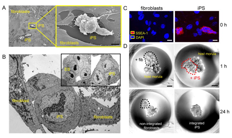
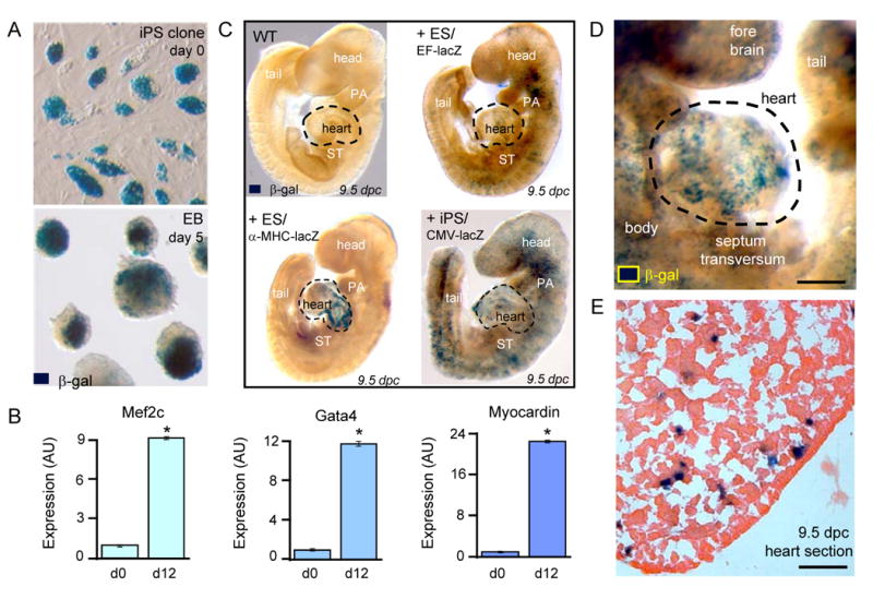
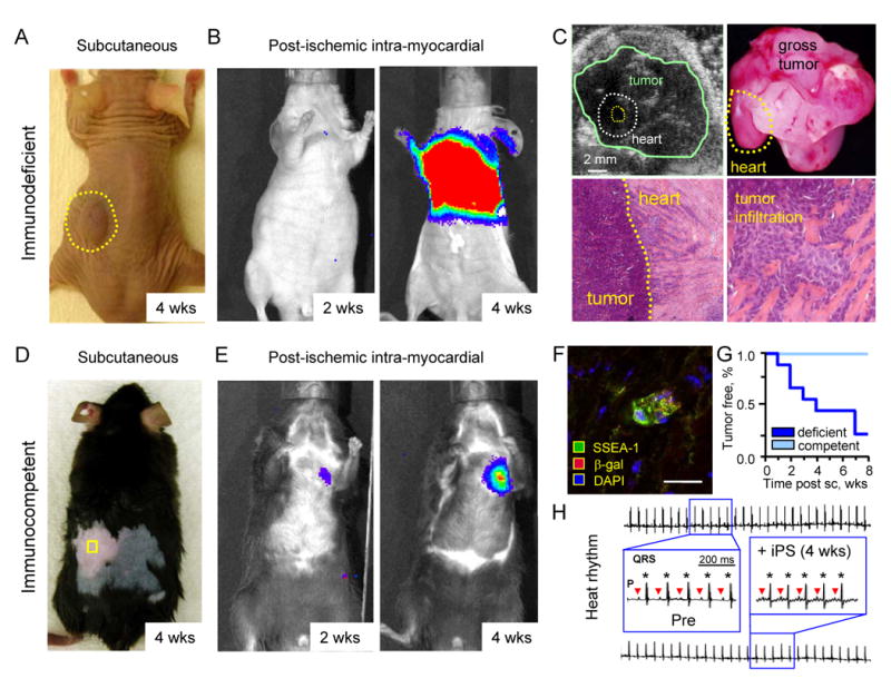
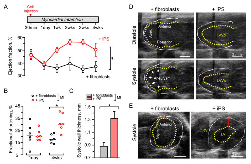
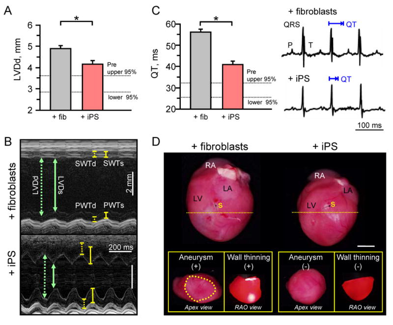
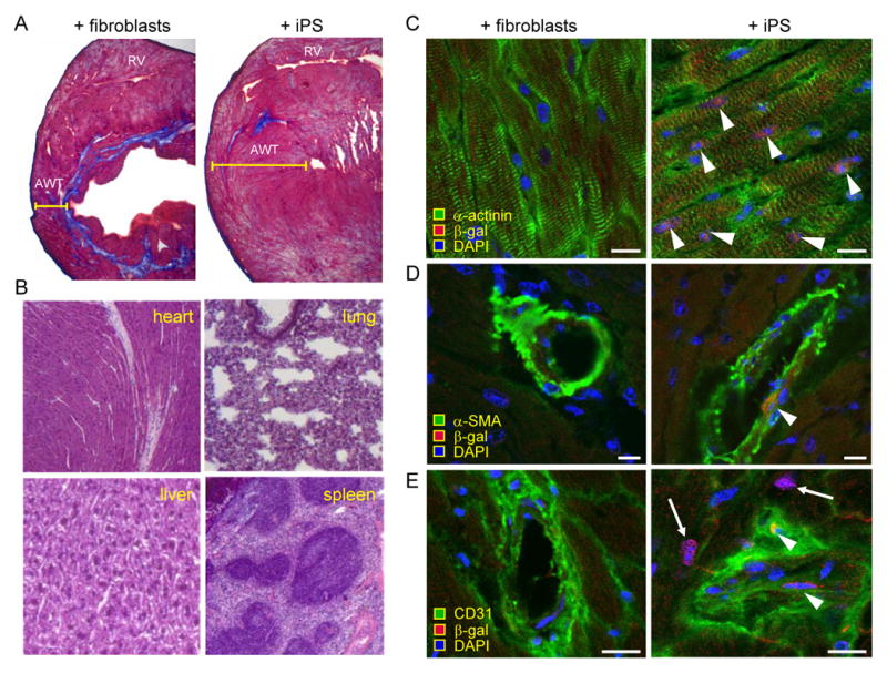
Methods and results: In this study, fibroblasts transduced with human stemness factors OCT3/4, SOX2, KLF4, and c-MYC converted into an embryonic stem cell-like phenotype and demonstrated the ability to spontaneously assimilate into preimplantation host morula via diploid aggregation, unique to bona fide pluripotent cells. In utero, iPS-derived chimera executed differentiation programs to construct normal heart parenchyma patterning. Within infarcted hearts in the adult, intramyocardial delivery of iPS yielded progeny that properly engrafted without disrupting cytoarchitecture in immunocompetent recipients. In contrast to parental nonreparative fibroblasts, iPS treatment restored postischemic contractile performance, ventricular wall thickness, and electric stability while achieving in situ regeneration of cardiac, smooth muscle, and endothelial tissue.

Conclusions: Fibroblasts reprogrammed by human stemness factors thus acquire the potential to repair acute myocardial infarction, establishing iPS in the treatment of heart disease.


Conflict of interest statement

Disclosures: None

Figures

Figure 1
Figure 1
Induced pluripotent stem cells (iPS) demonstrate pluripotent features. A, Flat fibroblasts reprogrammed with human stemness factors metamorphosed into rounded clusters shown by field-emission scanning electron microscopy. Bar=50 μm. B, In transmission electron microscopy, derived iPS demonstrated nuclear/cytoplasmic composition similar to embryonic stem cells (ES). C, Counterstained by nuclear DAPI, iPS expressed the pluripotent marker SSEA-1 (red), absent from fibroblasts (0 h; left). Bar=5 μm. D, Fibroblasts or iPS clumps were placed along with two 8-cell host embryos for diploid aggregation (1 h; top). Bar=30 μm. Within 24 h, iPS spontaneously integrated to form an early stage chimeric blastocyst (24 h; bottom right), in contrast to fibroblasts that were excluded (24 h; bottom left).
Figure 2
Figure 2
iPS recapitulate in utero cardiogenic propensity. A, LacZ-labeled iPS clones, detected by β-galactosidase (β-gal) staining, were maintained as undifferentiated colonies at day 0 before aggregation into embryoid bodies (EB). B, Gene expression profiles at day 0 (d0) compared to day 12 (d12) of differentiation demonstrated induction of cardiac transcription factors, Mef2c (p=0.049; n=3), Gata4 (p=0.049; n=3), and Myocardin (p=0.049; n=3). C, Embryos provide a wildtype (WT) environment to determine tissue-specific differentiation (upper left). Derived by diploid aggregations, ES stochastically contribute to tissue patterning with diffuse integration tracked with constitutively labelled EF-lacZ cell line (upper right) and cardiac-specific integration identified by α-MHC-lacZ reporter (lower left). iPS, labeled with ubiquitously expressing reporter with CMV promoter, identifies progeny throughout developing embryo (lower right). D, Chimerism with lacZ-labeled iPS demonstrated robust contribution to developing hearts within 9.5 dpc embryos. Bar=100 μm. E, Heart parenchyma of 9.5 dpc chimeric embryo contained integrated iPS progeny expressing β-galactosidase. Bar=50 μm.
Figure 3
Figure 3
iPS fate determined by host competency. A, Subcutaneous injection of 500,000 iPS in immunodeficient host resulted in tumor growth (dotted circle). B, Upon acute myocardial infarction, 200,000 iPS transplanted intra-myocardially were detected in the heart region by in vivo bioluminescence imaging dramatically expanding by 4 weeks (wks). C, Tumor growth was detected by echocardiography (upper left) and confirmed on necropsy in all immunodeficient hosts (upper right). Histology demonstrated tumor expansion outside of the heart (lower left), and infiltration within the wall of infarcted myocardium (lower right). D, Immunocompetent hosts reproducibly averted tumor growth upon subcutaneous injection (square) of 500,000 iPS throughout follow-up. E, iPS transplantation within infarcted myocardium of immunocompetent hosts produced stable engraftment detected by live-cell imaging throughout the 4 week follow-up. F, Post-ischemic myocardium transplanted with iPS at 4 weeks demonstrated rare pockets of SSEA-1 positive progeny. Bar=10 μm. G, Subcutaneous (sc) transplantation produced teratoma in immunodeficient (deficient), in contrast to tumor-free outcome in all immunocompetent (competent) hosts. H, Normal pre-infarction (Pre) sinus rhythm was maintained following iPS transplantation throughout the 4 week follow-up, with P-waves (triangles) preceding each QRS complex (stars) with no ventricular tachycardia or ectopy.
Figure 4
Figure 4
iPS restored function following acute myocardial infarction (MI). A, Upon randomization, cell-based intervention was performed at 30 min after coronary ligation. Divergent ejection fractions were noted in iPS (n=6) versus fibroblast (n=6) treated hearts within 1 week post-therapy, maintained throughout follow-up. *p=0.002 using two-way repeated measures ANOVA. B, Fractional shortening was similar at day 1 post-infarction, but significant improvement was only observed in iPS-treated hearts. Line indicates median value. *p=0.01. C, Septal wall thickness was preserved in systole following iPS (n=6) compared to fibroblast (n=6) treatment. *p=0.006. D, Echocardiography with long-axis views revealed anterior wall thinning and apex aneurysmal formation (arrow heads) in fibroblast-treated hearts as indicated by akinetic wall (left) in contrast to normalized systolic wall motion in iPS-treated hearts (right). E, Short-axis confirmed thinning in the anterior wall (red bar) and overall decreased cardiac performance with fibroblast compared to iPS-based interventions. Yellow and white dotted lines indicate endocardium and epicardium, respectively. LVVd: left ventricular volume in diastole; LVVs: left ventricular volume in systole.
Figure 5
Figure 5
iPS halt maladaptive remodeling and preserve structure. A, Diastolic parameters revealed a significant decrease in global left ventricular diastolic diameter (LVDd) in hearts treated with iPS (n=6) compared to fibroblasts (n=6) at 4-weeks post-therapy (*p=0.007). B, M-mode echocardiography demonstrated dilated ventricular lumen with reduced anterior and septal wall thickness (SWTd) during systole in fibroblast-treated hearts (upper), which improved with iPS intervention (lower). C, Time required for ventricular repolarization and depolarization, measured by the QT interval, was significantly prolonged in fibroblast (n=6) compared to iPS (n=6) treated hearts. *p=0.004. D, Hearts were pathologically enlarged in the fibroblast-treated group with aneurysmal formation (+) and severe wall thinning (+) visible with translumination compared to structurally preserved iPS-treated hearts with normal apex geometry (-) and opaque thick walls (-) on right anterior-oblique (RAO) view upon transverse sectioning of hearts immediately inferior to the site of surgical ligation (yellow dotted line). Bar=5mm. Aneurysm delineated by yellow dotted circle. RA: right atrium; LA: left atrium; LV: left ventricle; s: suture; SWTd: septal wall thickness in diastole; SWTs: septal wall thickness in systole; PWTd: posterior wall thickness in diastole; PWTs: posterior wall thickness in systole.
Figure 6
Figure 6
iPS treatment reduced scar and contributed to multi-lineage reconstruction. A, After 4 week of therapy, Masson's trichrome staining demonstrated reduced anterior wall thickness (AWT) and fibrosis (blue staining) in hearts treated with fibroblasts (left) compared to iPS (right). B, Autopsy demonstrated tumor-free heart, liver, lung, or spleen in the iPS-treated cohort. C, After 4 weeks, integrated iPS progeny expressed markers of remuscularization according to α-actinin (right) and β-gal co-expression (arrow heads), not detectable with fibroblast treatment (left). D, Smooth muscle actin (α-SMA; arrow head), and E, CD31 positive endothelium (arrow heads) were identified in iPS progeny (right) compared to no expression with fibroblast treatment (left). DAPI visualized nuclei. Bar=5 μm.

References

    1. Daley GQ, Scadden DT. Prospects for stem cell-based therapy. Cell. 2008;132:544–548. - PubMed
    1. Segers V, Lee RT. Stem-cell therapy for cardiac disease. Nature. 2008;451:937–942. - PubMed
    1. Yamanaka S. Strategies and new developments in the generation of patient-specific pluripotent stem cells. Cell Stem Cell. 2007;1:39–49. - PubMed
    1. Nishikawa S, Goldstein RA, Nierras CR. The promise of human induced pluripotent stem cells for research and therapy. Nat Rev Mol Cell Biol. 2008;9:725–729. - PubMed
    1. Takahashi K, Okita K, Nakagawa M, Yamanaka S. Induction of pluripotent stem cells from mouse embryonic and adult fibroblast cultures by defined factors. Cell. 2006;126:663–676. - PubMed

Publication types

MeSH terms