

Using dermoscopic criteria and patient-related factors for the management of pigmented melanocytic nevi
- PMID: 19620566
- PMCID: PMC2856040
- DOI: 10.1001/archdermatol.2009.115
Using dermoscopic criteria and patient-related factors for the management of pigmented melanocytic nevi
Abstract
Objective: To review recent dermoscopy studies that provide new insights into the evolution of nevi and their patterns of pigmentation as they contribute to the diagnosis of nevi and the management of pigmented melanocytic nevi.
Data sources: Data for this article were identified by searching the English and German literature by Medline and Journals@Ovid search for the period 1950 to January 2009.
Study selection: The following relevant terms were used: dermoscopy, dermatoscopy, epiluminescence microscopy (ELM), surface microscopy, digital dermoscopy, digital dermatoscopy, digital epiluminescence microscopy, digital surface microscopy, melanocytic skin lesion, nevi, and pigmented skin lesions. There were no exclusion criteria.
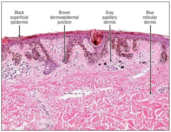
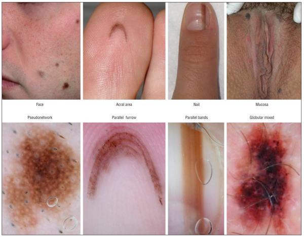
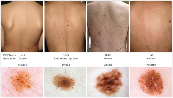
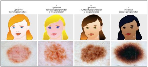
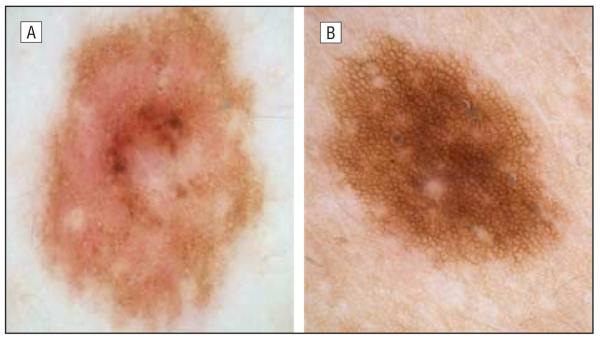
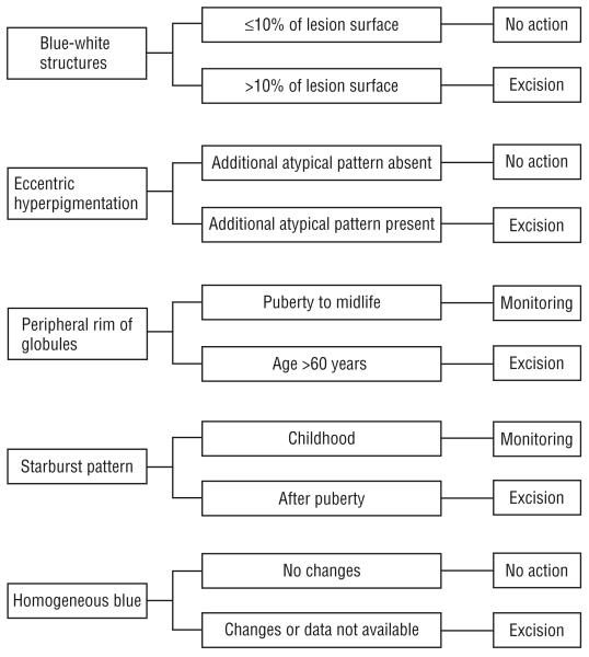
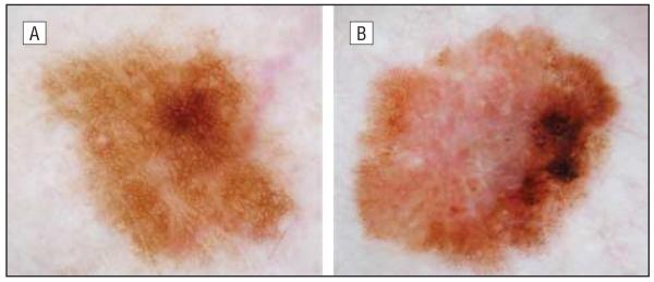
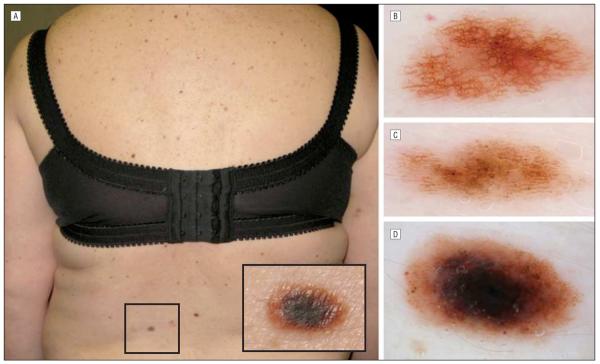
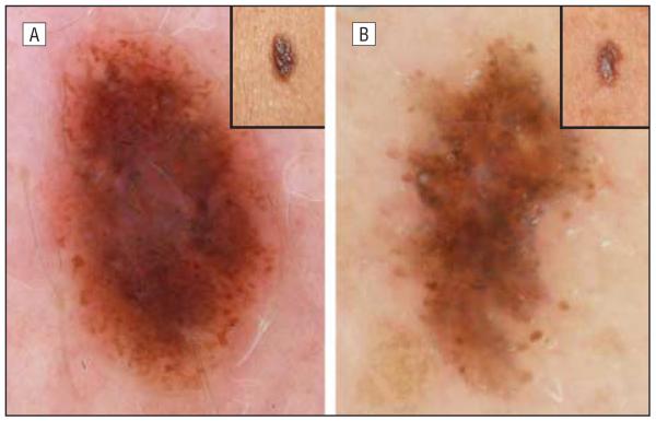
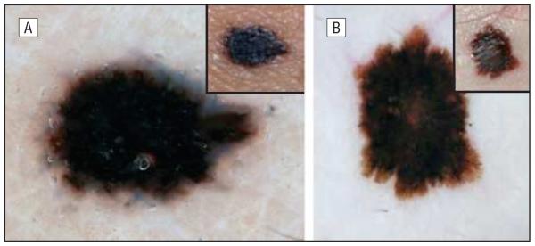
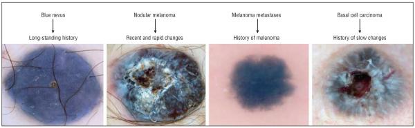
Data synthesis: The dermoscopic diagnosis of nevi relies on the following 4 criteria (each of which is characterized by 4 variables): (1) color (black, brown, gray, and blue); (2) pattern (globular, reticular, starburst, and homogeneous blue pattern); (3) pigment distribution (multifocal, central, eccentric, and uniform); and (4) special sites (face, acral areas, nail, and mucosa). In addition, the following 6 factors related to the patient might influence the pattern of pigmentation of the individual nevi: age, skin type, history of melanoma, UV exposure, pregnancy, and growth dynamics.
Conclusions: The 4 x 4 x 6 "rule" may help clinicians remember the basic dermoscopic criteria of nevi and the patient-related factors influencing their patterns. Dermoscopy is a useful technique for diagnosing melanocytic nevi, but the clinician should take additional factors into consideration to optimize the management of cases of pigmented lesions.
Figures

References
-
- Bystryn JC. Epiluminescence microscopy: a reevaluation of its purpose. Arch Dermatol. 2001;137(3):377–379. - PubMed
-
- Carli P, De Giorgi V, Crocetti E, et al. Improvement of malignant/benign ratio in excised melanocytic lesions in the “dermoscopy era”: a retrospective study 1997-2001. Br J Dermatol. 2004;150(4):687–692. - PubMed
-
- Gachon J, Beaulieu P, Sei JF, et al. First prospective study of the recognition process of melanoma in dermatological practice. Arch Dermatol. 2005;141(4):434–438. - PubMed
-
- Scope A, Dusza SW, Halpern AC, et al. The “ugly duckling” sign: agreement between observers. Arch Dermatol. 2008;144(1):58–64. - PubMed
-
- Hofmann-Wellenhof R, Blum A, Wolf IH, et al. Dermoscopic classification of atypical melanocytic nevi (Clark nevi) Arch Dermatol. 2001;137(12):1575–1580. - PubMed
Publication types
MeSH terms
Grants and funding
LinkOut - more resources
Full Text Sources
Other Literature Sources
Medical
