The selectivity and functional connectivity of the anterior temporal lobes
- PMID: 19620621
- PMCID: PMC2837089
- DOI: 10.1093/cercor/bhp149
The selectivity and functional connectivity of the anterior temporal lobes
Abstract
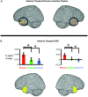
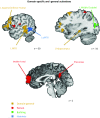
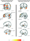
One influential account asserts that the anterior temporal lobe (ATL) is a domain-general hub for semantic memory. Other evidence indicates it is part of a domain-specific social cognition system. Arbitrating these accounts using functional magnetic resonance imaging has previously been difficult because of magnetic susceptibility artifacts in the region. The present study used parameters optimized for imaging the ATL, and had subjects encode facts about unfamiliar people, buildings, and hammers. Using both conjunction and region of interest analyses, person-selective responses were observed in both the left and right ATL. Neither building-selective, hammer-selective nor domain-general responses were observed in the ATLs, although they were observed in other brain regions. These findings were supported by "resting-state" functional connectivity analyses using independent datasets from the same subjects. Person-selective ATL clusters were functionally connected with the brain's wider social cognition network. Rather than serving as a domain-general semantic hub, the ATLs work in unison with the social cognition system to support learning facts about others.
Figures


Similar articles
-
Differential contributions of the anterior temporal and medial temporal lobe to the retrieval of memory for person identity information.Hum Brain Mapp. 2008 Dec;29(12):1343-54. doi: 10.1002/hbm.20469. Hum Brain Mapp. 2008. PMID: 17948885 Free PMC article.
-
Sensory and semantic category subdivisions within the anterior temporal lobes.Neuropsychologia. 2011 Oct;49(12):3419-29. doi: 10.1016/j.neuropsychologia.2011.07.033. Epub 2011 Aug 22. Neuropsychologia. 2011. PMID: 21889520 Free PMC article.
-
Dissociable roles of the anterior temporal regions in successful encoding of memory for person identity information.J Cogn Neurosci. 2010 Oct;22(10):2226-37. doi: 10.1162/jocn.2009.21349. J Cogn Neurosci. 2010. PMID: 19803684
-
Graded specialization within and between the anterior temporal lobes.Ann N Y Acad Sci. 2015 Nov;1359(1):84-97. doi: 10.1111/nyas.12951. Epub 2015 Oct 26. Ann N Y Acad Sci. 2015. PMID: 26502375 Free PMC article. Review.
-
Is the difference between right and left ATLs due to the distinction between general and social cognition or between verbal and non-verbal representations?Neurosci Biobehav Rev. 2015 Apr;51:296-312. doi: 10.1016/j.neubiorev.2015.02.004. Epub 2015 Feb 16. Neurosci Biobehav Rev. 2015. PMID: 25697904 Review.
Cited by
-
Looking beyond the face area: lesion network mapping of prosopagnosia.Brain. 2019 Dec 1;142(12):3975-3990. doi: 10.1093/brain/awz332. Brain. 2019. PMID: 31740940 Free PMC article.
-
Intracerebral Electrophysiological Recordings to Understand the Neural Basis of Human Face Recognition.Brain Sci. 2023 Feb 18;13(2):354. doi: 10.3390/brainsci13020354. Brain Sci. 2023. PMID: 36831897 Free PMC article. Review.
-
Face Processing in Developmental Prosopagnosia: Altered Neural Representations in the Fusiform Face Area.Front Behav Neurosci. 2021 Nov 18;15:744466. doi: 10.3389/fnbeh.2021.744466. eCollection 2021. Front Behav Neurosci. 2021. PMID: 34867227 Free PMC article.
-
What's unique about unique entities? An fMRI investigation of the semantics of famous faces and landmarks.Cereb Cortex. 2012 Sep;22(9):2005-15. doi: 10.1093/cercor/bhr274. Epub 2011 Oct 20. Cereb Cortex. 2012. PMID: 22021913 Free PMC article.
-
Semantic variability predicts neural variability of object concepts.Neuropsychologia. 2015 Sep;76:41-51. doi: 10.1016/j.neuropsychologia.2014.11.029. Epub 2014 Nov 25. Neuropsychologia. 2015. PMID: 25462197 Free PMC article.
References
-
- Addis DR, McAndews MP. Prefrontal and hippocampal contributions to the generation and binding of semantic associations during successful encoding. Neuroimage. 2006;33:1194–1206. - PubMed
-
- Amodio DM, Frith CD. Meeting of minds: the medial frontal cortex and frontal cognition. Nat Rev Neurosci. 2006;7:268–277. - PubMed
-
- Badre D, Wagner AD. Left ventrolateral prefrontal cortex and the cognitive control of memory. Neuropsychologia. 2007;45:2883–2901. - PubMed
-
- Baldo JV, Shimamura AP. Letter and category fluency in patients with frontal lobe lesions. Neuropsychology. 1998;12:259–267. - PubMed
-
- Barsalou LW. Perceptual symbol systems. Behav Brain Sci. 1999;22:577–609. - PubMed
Publication types
MeSH terms
Substances
LinkOut - more resources
Full Text Sources
Molecular Biology Databases

