. 2009 Oct;8(10):2308-20.
doi: 10.1074/mcp.M900178-MCP200. Epub 2009 Jul 20.

A mediator of Rho-dependent invasion moonlights as a methionine salvage enzyme

Affiliations

A mediator of Rho-dependent invasion moonlights as a methionine salvage enzyme

Yukihito Kabuyama et al. Mol Cell Proteomics. 2009 Oct.

Abstract

RhoA controls changes in cell morphology and invasion associated with cancer phenotypes. Cell lines derived from melanoma tumors at varying stages revealed that RhoA is selectively activated in cells of metastatic origin. We describe a functional proteomics strategy to identify proteins regulated by RhoA and report a previously uncharacterized human protein, named "mediator of RhoA-dependent invasion (MRDI)," that is induced in metastatic cells by constitutive RhoA activation and promotes cell invasion. In human melanomas, MRDI localization correlated with stage, showing nuclear localization in nevi and early stage tumors and cytoplasmic localization with plasma membrane accentuation in late stage tumors. Consistent with its role in promoting cell invasion, MRDI localized to cell protrusions and leading edge membranes in cultured cells and was required for cell motility, tyrosine phosphorylation of focal adhesion kinase, and modulation of actin stress fibers. Unexpectedly MRDI had enzymatic function as an isomerase that converts the S-adenosylmethionine catabolite 5-methylribose 1-phosphate into 5-methylribulose 1-phosphate. The enzymatic function of MRDI was required for methionine salvage from S-adenosylmethionine but distinct from its function in cell invasion. Thus, mechanisms used by signal transduction pathways to control cell movement have evolved from proteins with ancient function in amino acid metabolism.


Figures

Fig. 1.
Fig. 1.
Stage-specific activation of RhoA in melanoma cells. RhoA activation was measured by Rho-GTP binding to GST-RBD. Western blots show levels of RhoA-GTP bound to GST-RBD in pulldown assays and RhoA and γ-tubulin in total cell lysates. Similar results were observed in three independent experiments. RGP, radial growth phase; VGP, vertical growth phase; M'cyte, melanocyte.
Fig. 2.
Fig. 2.
A functional proteomics screen for RhoA targets. A, outline of the experimental strategy for identifying proteins responsive to RhoA. Step 1, protein responses to RhoA-V14 are profiled in WM35 cells by 2DE to identify pathway targets. Step 2, these targets are examined in other melanoma cell lines to identify those that correlate with RhoA activation. B, sequence of human MGC3207 indicating peptides observed by peptide mass matching (underlined) or MS/MS sequencing (boxed). C, 2DE images showing MGC3207 protein changes responsive to RhoA in WM35 cells and across melanocyte and melanoma cell lines. Arrowheads show protein intensities that are unaltered (open) or altered in the same direction as those induced by RhoA signaling (closed). RGP, radial growth phase; VGP, vertical growth phase.
Fig. 3.
Fig. 3.
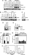
MRDI is induced by RhoA and mediates RhoA-dependent cell invasion. A–E, Western blots demonstrate that RhoA induces expression of MRDI. A, blots probed with anti-MRDI show elevated protein levels in metastatic cell lines with constitutive RhoA activity. RhoA-GTP blots are from the image in Fig. 1A. Loading controls show α-tubulin in total lysates. B, RhoA is necessary for MRDI induction. Extracts (5 μg) were probed with anti-MRDI, examining WM35 cells treated by adenoviral delivery of Ad-CMV, Ad-Rac1-V12, or Ad-RhoA-V14 or pretreated with Y27632 prior to Ad-RhoA-V14 (lanes 1–4). A375 cells were treated with Ad-CMV, Ad-DN-Rac1, C3 transferase, or Y27632 (lanes 5–8). C, A375 cells were treated with Ad-CMV or Ad-DN-RhoA-N19, and lysates were blotted with anti-MRDI. D, A375 cells were treated with RNAi for 48 h to inhibit expression of RhoA or RhoC, and lysates were blotted with anti-MRDI. Anti-RhoA or anti-RhoC Westerns demonstrate selective knockdown by RNAi. E, control or RhoC-expressing WM35 cells were examined for expression of MRDI and RhoC, and RhoC-GTP was assayed by GST-rhotekin pulldown assays. RhoC overexpression and activation had little effect on MRDI compared with cells treated with lysophosphatidic acid (LPA) (24 h). F–H, MRDI promotes cell invasion. F, RNAi knockdown of MRDI in metastatic melanoma A375 and HS294T cells inhibits cell invasion through Matrigel. G, invasion is suppressed in A375 cells treated with four different MRDI-RNAi oligonucleotide sequences, singly or in combination, but not in cells treated with lamin A/C-RNAi (48-h transfection). Values represent mean ± S.D. (n = 3). H, cell invasion depends on Rho signaling. Invasion is reduced in A375 cells treated with RhoA-RNAi, RhoC-RNAi, or C3 transferase. RGP, radial growth phase; VGP, vertical growth phase.
Fig. 4.
Fig. 4.
MRDI localizes to membranes and cell protrusions. A and B, MRDI localization correlates with tumor grade in human melanoma specimens. A, junctional compound nevus shows nuclear localization but little cytoplasmic immunoreactivity. B, melanoma in situ shows MRDI immunoreactivity localized to cytoplasmic pools. The inset at higher magnification illustrates membrane accentuation of immunoreactivity (arrowheads). C, MRDI immunostaining shows membrane accentuation between cells in xenograft tumors of A375 cells grown in athymic nude mice (arrowheads). D, MRDI localizes to cell protrusions in cultured A375 cells when probing endogenous MRDI with anti-MRDI (1:3000). Cells treated with MRDI-RNAi show loss of reactivity at the membrane leading edge and protrusions and partial loss of nuclear staining. Western blots of lysates (5 μg) probed with anti-MRDI confirm knockdown (left panel). E, RPE cells transfected with MRDI or empty vector and probed with anti-MRDI antibody (Ab) and CM-DiIC18 (CM-dil) show localization of MRDI at the membrane periphery and leading edge. Western blots of lysates (20 μg) show low protein expression in untransfected cells and elevated expression upon transfection (left panel). F, indirect immunofluorescence of RPE cells shows colocalization of MRDI with N-WASP at the membrane leading edge.
Fig. 5.
Fig. 5.
MRDI modulates cell motility, FAK phosphorylation, and stress fiber formation. A, live cell images of A375 cells treated with control scrambled oligonucleotide or MRDI-RNAi show decreased cell movement following MRDI knockdown. Images are taken at t = 0 after serum addition, and traces show nuclear movement over the next 60 min. Right panel, summary of several biological replicates quantifying percentages of control and MRDI-RNAi cells that move by less than one (“0”), between one and two (“1”), and more than two (“2”) nuclear diameters in 60 min. Values show mean ± S.E. (number of experiments, n = 9 control and n = 8 RNAi). B, FAK-Tyr(P)397 is up-regulated in WM35 cells expressing Ad-RhoA-V14 (lanes 1 and 2) and inhibited in A375 cells treated with C3 transferase (lanes 3 and 4) or MRDI-RNAi (lanes 5 and 6). In contrast, total FAK protein expression is unaffected. C, MRDI knockdown enhances actin polymerization. A375 cells were transfected for 48 h with or without MRDI-RNAi and observed following phalloidin staining. Two representative cells are shown in each experiment.
Fig. 6.
Fig. 6.
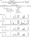
MRDI is a methylthioribose-1-phosphate isomerase. A, enzymatic steps in the methionine salvage pathway. Following conversion of decarboxylated S-AdoMet to MTA during polyamine synthesis, MTA is catabolized to methionine. B–E, recombinant purified MRDI shows MTR-1-P isomerase enzymatic activity. 1H NMR spectra, collected after in vitro reactions in the absence (B) or presence (C) of wild type MRDI, show that MRDI catalyzes the conversion of MTR-1-P to MTRu-1-P. In contrast, catalytic turnover is absent in reactions with MRDI-C168S (D) or MRDI-D248A (E) mutants. Peak assignments (a–f) in B and C correspond to protons indicated in A.
Fig. 7.
Fig. 7.
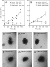
Catalytic activity of MRDI is required for methionine salvage but not cell invasion. A, A375 cells grown in the absence of methionine (▲) show growth arrest due to lack of an essential amino acid in the medium. This is partially rescued by incubation with 100 μm MTA in control cells (●). MTA fails to rescue growth in cells in which MRDI expression is stably inhibited by shRNA (♢), demonstrating a requirement for MRDI in methionine salvage. B, MRDI-shRNA knockdown cells were grown in the presence of MTA and the absence of methionine. Growth was rescued by add-back expression of MRDI-WT (♦) but not C168S (■) or D248A (○) mutants, indicating that MRDI catalytic activity is needed for cell growth via methionine salvage. C–H, A375 cells grown as spheroids show suppression of invasion into 3D collagen matrix upon stable knockdown of MRDI with shRNA. The invasion phenotype is rescued equally well by MRDI-WT, -C168S, and -D248A, indicating that catalytic activity is not required for cell invasion.

Similar articles

Cited by

References

    1. Bogenrieder T., Herlyn M. (2002) Cell-surface proteolysis, growth factor activation and intercellular communication in the progression of melanoma. Crit. Rev. Oncol. Hematol 44, 1–15 - PMC - PubMed
    1. Sauter E. R., Herlyn M. (1998) Molecular biology of human melanoma development and progression. Mol. Carcinog 23, 132–143 - PubMed
    1. Jaffe A. B., Hall A. (2005) Rho GTPases: biochemistry and biology. Annu. Rev. Cell Dev. Biol 21, 247–269 - PubMed
    1. Clark E. A., Golub T. R., Lander E. S., Hynes R. O. (2000) Genomic analysis of metastasis reveals an essential role for RhoC. Nature 406, 532–535 - PubMed
    1. Ikoma T., Takahashi T., Nagano S., Li Y. M., Ohno Y., Ando K., Fujiwara T., Fujiwara H., Kosai K. (2004) A definitive role of RhoC in metastasis of orthotopic lung cancer in mice. Clin. Cancer Res 10, 1192–1200 - PubMed

Publication types

MeSH terms