. 2009 Aug 3;206(8):1739-53.
doi: 10.1084/jem.20090004. Epub 2009 Jul 20.

Pre-B cell receptor-mediated cell cycle arrest in Philadelphia chromosome-positive acute lymphoblastic leukemia requires IKAROS function

Affiliations

Pre-B cell receptor-mediated cell cycle arrest in Philadelphia chromosome-positive acute lymphoblastic leukemia requires IKAROS function

Daniel Trageser et al. J Exp Med. .

Abstract

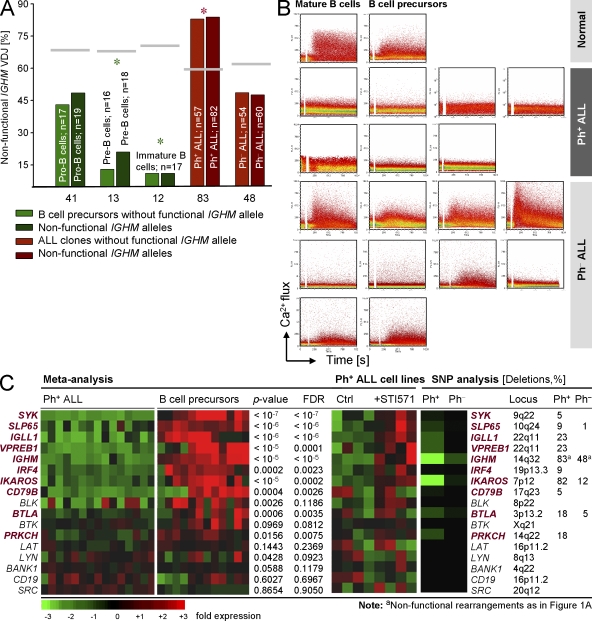
B cell lineage acute lymphoblastic leukemia (ALL) arises in virtually all cases from B cell precursors that are arrested at pre-B cell receptor-dependent stages. The Philadelphia chromosome-positive (Ph(+)) subtype of ALL accounts for 25-30% of cases of adult ALL, has the most unfavorable clinical outcome among all ALL subtypes and is defined by the oncogenic BCR-ABL1 kinase and deletions of the IKAROS gene in >80% of cases. Here, we demonstrate that the pre-B cell receptor functions as a tumor suppressor upstream of IKAROS through induction of cell cycle arrest in Ph(+) ALL cells. Pre-B cell receptor-mediated cell cycle arrest in Ph(+) ALL cells critically depends on IKAROS function, and is reversed by coexpression of the dominant-negative IKAROS splice variant IK6. IKAROS also promotes tumor suppression through cooperation with downstream molecules of the pre-B cell receptor signaling pathway, even if expression of the pre-B cell receptor itself is compromised. In this case, IKAROS redirects oncogenic BCR-ABL1 tyrosine kinase signaling from SRC kinase-activation to SLP65, which functions as a critical tumor suppressor downstream of the pre-B cell receptor. These findings provide a rationale for the surprisingly high frequency of IKAROS deletions in Ph(+) ALL and identify IKAROS-mediated cell cycle exit as the endpoint of an emerging pathway of pre-B cell receptor-mediated tumor suppression.


Figures

Figure 1.
Figure 1.
Pre-B cell receptor function in normal human B cell precursors and Ph+ ALL. The configuration of the Ig heavy chain (IGHM) locus was studied in bone marrow pro–B cells, pre–B cells, immature B cells, 57 cases of Ph+ ALL, and 54 cases of Ph-negative ALL (A). The frequencies of cells/ALL clones lacking a functional IGHM gene rearrangement (light bars) and the total frequency of nonfunctional IGHM alleles in these populations (dark bars) are shown. The expected frequencies of cells/ ALL clones lacking coding capacity for a pre–B cell receptor based on random distribution (Table S1) are indicated as horizontal gray lines. Asterisks denote significant differences from random distribution (P < 0.05). Ca2+ mobilization in response to pre–B cell receptor engagement was studied in normal B and pre–B cells, 7 cases of Ph+ ALL and 10 Ph-negative ALL (B). A metaanalysis of published gene expression data for pre–B cell receptor–related genes in 15 cases of Ph+ ALL and normal human B cell precursors was performed (C, left). P values and false discovery rates (FDR) are indicated. Ph+ ALL cell lines (BV173, Nalm1, SUP-B15, and TOM1) were cultured in the presence or absence of 10 μmol/STI571 (Imatinib) for 16 h and analyzed by Affymetrix U133A2.0 microarrays (C, middle). 22 cases of Ph+ ALL were analyzed by a SNP mapping assay (C, right). The frequency of deletions is given in percent. The frequencies of deletions in our dataset of 22 cases of Ph+ ALL are plotted against the frequencies of deletions found in Ph ALL (Mullighan et al., 2007).
Figure 2.
Figure 2.
Progressive leukemic transformation of B cell precursors in BCR-ABL1 transgenic mice. B cell precursors from wild-type and BCR-ABL1 transgenic mice were analyzed for their clonality using spectratyping of Ig gene rearrangements (A), μ chain expression (B), pre–B cell receptor responsiveness (C), and gene expression pattern (D), and RT-PCR validation (E; three experiments). Data in A–C are representative of results from six independent experiments with at least two mice per group. B cell precursor populations were compared from wild-type animals, preleukemic BCR-ABL1 transgenic mice (age <60 d), BCR-ABL1 transgenic mice with full-blown leukemia (age >90 d), and after treatment of mice with AMN107 (75 mg/kg/d) for 7 d.
Figure 3.
Figure 3.
Reconstitution of Slp65 deficiency in BCR-ABL1 ALL cells suppresses leukemic growth BCR-ABL1–transformed Slp65−/−. ALL cells were reconstituted with Slp65/GFP or a GFP empty vector control (A; single measurements in three independent experiments). For inducible Slp65 reconstitution, we fused Slp65 to the estrogen receptor ligand-binding domain (ERT2). Either ERT2 fused to the N terminus of Slp65 (ERT2-Slp65/GFP) or ERT2 alone (ERT2/GFP) were expressed in the Slp65−/− BCR-ABL1–transformed ALL cells and activated by addition of 1 μmol/l 4-hydroxy-tamoxifen (OHT) using ethanol (EtOH) as vehicle control (B; triplicate measurements, the experiment was repeated once). Primary human leukemia cells from one case of Ph+ ALL cells lacking expression of SLP65 were cultured on OP9 stroma in the presence of IL-7 and transduced with SLP65/GFP or a GFP empty vector control (three independent transductions) and monitored by flow cytometry (C). BCR-ABL1–transformed Slp65−/− ALL cells were labeled by lentiviral firefly luciferase, transduced with retroviral vectors encoding either Slp65/GFP or GFP alone and injected into five sublethally irradiated NOD/SCID mice per group. Engraftment and leukemic growth was monitored by luciferase-bioimaging (D; experiment repeated once). The blue scale bar in (D) corresponds to 1 cm in length. Leukemic infiltration (CD19+ GFP+) of bone marrow, spleen, and peripheral blood (three independent experiments) was documented by flow cytometry (E; representative data from two experiments).
Figure 4.
Figure 4.
Reconstitution of μ chain expression results in leukemia suppression BCR-ABL1–transformed Ighm−/−. ALL cells were transduced with retroviral expression vectors encoding either CD8 alone or CD8 with a functional μ heavy chain. Enrichment or depletion of CD8+ and μ/CD8+ cells was monitored over 10 d (A; three independent transductions). Primary human Ph+ ALL cells lacking coding capacity for expression of a μ chain were cultured on OP9 stroma in the presence of IL-7 and transduced with μ chain/CD8 or CD8 alone and monitored by flow cytometry (B; three independent transductions). BCR-ABL1 transgenic mouse B cell lineage leukemia cells lacking μ chain expression were transduced with retroviral expression vectors encoding either CD8 alone or CD8/μ chain (C and D). Percentages of Annexin V+ and propidium iodide+ cells were determined after 2 and 5 d of transduction and means of three experiments are indicated in (D).
Figure 5.
Figure 5.
Reconstitution of pre–B cell receptor signaling in BCR-ABL1 ALL cells induces cell cycle arrest. Rag2−/− tTA/μ chain transgenic mice are unable to express an endogenous μ chain but carry a functionally prerearranged μ chain under control of Tet operator (tetO) sequences. These mice express a tTA under control of endogenous μ chain regulatory elements and withdrawal of Tet results in activation of μ chain expression (A; routinely performed quality control). The effect of Tet-inducible activation of μ chain expression in BCR-ABL1–transformed ALL cells on leukemic growth was measured in a detailed cell cycle analysis (B-C). Percentages of cells in G0/G1, S and G2/M phases of the cell cycle were calculated and means of three experiments are indicated in C.
Figure 6.
Figure 6.
Pre-B cell receptor–mediated suppression of leukemic growth requires IKAROS function. mRNA levels of Ikaros after μ chain induction were measured by quantitative RT-PCR (A; three experiments). BCR-ABL1–transformed ALL cells were retrovirally transduced with the dominant-negative IKAROS splice variant IK6/GFP or a GFP empty vector control (B–D). Leukemia cells transduced with IK6/GFP (+IK6) or an empty vector control (-IK6) were studied in the presence or absence of pre–B cell receptor activation (+/− μ chain). IK6/GFP- and GFP-transduced leukemia cells were subjected to cell cycle analysis in the presence or absence of inducible pre–B cell receptor signaling (B–D). The percentages of cells in G0/G1, S and G2/M phase are indicated and means of three experiments are indicated in C. In D, the relative increase or decrease of IK6/GFP+ versus GFP+ leukemia cells in the presence or absence of pre–B cell receptor activation (± μ chain) was measured. The experiment was repeated once.
Figure 7.
Figure 7.
Reconstitution of IKAROS expression in Ph+ ALL cells results in BCR-ABL1-mediated activation of SLP65 even in the absence of a pre–B cell receptor. Genotype and expression of IGHM (μ chain), SLP65, and IKAROS (both functional IK1 and dominant-negative IK6 forms of IKAROS) were studied in three cases of Ph+ ALL. Ph+ ALL cells were transduced once in each case with retroviral vectors encoding SLP65/GFP, IKAROS (IK1)/GFP and μ chain/CD8 or GFP- and CD8-empty vector controls. Enrichment or depletion of GFP+ and CD8+ cells was monitored by flow cytometry (A; three cases were studied). IKAROS-deficient leukemia cells from Ph+ ALL cases 1 and 2 were transduced with a retroviral expression vector for IKAROS (IK1)/GFP and a GFP empty vector control. 2 d after transduction, GFP+ cells were sorted and cultured in the presence or absence of 10 μmol/liter Imatinib for 12 h (B). Protein lysates from these cells were then subjected to Western blot analysis using antibodies against the SRC family kinase LYN, activated SRC kinases (including LYN) that are phosphorylated at Y416, the ubiquitin-ligase CBL, activated CBL phosphorylated at Y731, SLP65, and activated SLP65 phosphorylated at Y96. Antibodies against EIF4E were used as a loading control. Experiments on two cases were performed. In (C), pre–B cells isolated from bone marrow of Slp65−/− mice were transformed by a retrovirus encoding BCR-ABL1/Neo. In three experiments, BCR-ABL1–transformed Slp65−/− ALL cells were reconstituted with retroviral expression vectors encoding wild-type SLP65/GFP or the mutant SLP65Y96F/GFP or transduced with a GFP empty vector control (C).

References

    1. Bäckesjö C.M., Vargas L., Superti-Furga G., Smith C.I. 2002. Phosphorylation of Bruton's tyrosine kinase by c-Abl.Biochem. Biophys. Res. Commun. 299:510–515 - PubMed
    1. Cheng A.M., Rowley B., Pao W., Hayday A., Bolen J.B., Pawson T. 1995. Syk tyrosine kinase required for mouse viability and B-cell development.Nature. 378:303–306 - PubMed
    1. Danial N.N., Pernis A., Rothman P.B. 1995. Jak-STAT signaling induced by the v-abl oncogene.Science. 269:1875–1877 - PubMed
    1. Feldhahn N., Klein F., Mooster J.L., Hadweh P., Sprangers M., Wartenberg M., Bekhite M.M., Hofmann W.K., Herzog S., Jumaa H., et al. 2005. Mimicry of a constitutively active pre-B cell receptor in acute lymphoblastic leukemia cells.J. Exp. Med. 201:1837–1852 - PMC - PubMed
    1. Flemming A., Brummer T., Reth M., Jumaa H. 2003. The adaptor protein SLP65 acts as a tumor suppressor that limits pre-B cell expansion.Nat. Immunol. 4:38–43 - PubMed

Publication types

MeSH terms

Substances