An activating mutation in the CSF3R gene induces a hereditary chronic neutrophilia
- PMID: 19620628
- PMCID: PMC2722170
- DOI: 10.1084/jem.20090693
An activating mutation in the CSF3R gene induces a hereditary chronic neutrophilia
Abstract
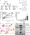
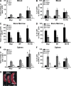
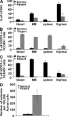
We identify an autosomal mutation in the CSF3R gene in a family with a chronic neutrophilia. This T617N mutation energetically favors dimerization of the granulocyte colony-stimulating factor (G-CSF) receptor transmembrane domain, and thus, strongly promotes constitutive activation of the receptor and hypersensitivity to G-CSF for proliferation and differentiation, which ultimately leads to chronic neutrophilia. Mutant hematopoietic stem cells yield a myeloproliferative-like disorder in xenotransplantation and syngenic mouse bone marrow engraftment assays. The survey of 12 affected individuals during three generations indicates that only one patient had a myelodysplastic syndrome. Our data thus indicate that mutations in the CSF3R gene can be responsible for hereditary neutrophilia mimicking a myeloproliferative disorder.
Figures


Similar articles
-
Gain-of-function mutations in granulocyte colony-stimulating factor receptor (CSF3R) reveal distinct mechanisms of CSF3R activation.J Biol Chem. 2018 May 11;293(19):7387-7396. doi: 10.1074/jbc.RA118.002417. Epub 2018 Mar 23. J Biol Chem. 2018. PMID: 29572350 Free PMC article.
-
The CSF3R T618I mutation causes a lethal neutrophilic neoplasia in mice that is responsive to therapeutic JAK inhibition.Blood. 2013 Nov 21;122(22):3628-31. doi: 10.1182/blood-2013-06-509976. Epub 2013 Sep 30. Blood. 2013. PMID: 24081659 Free PMC article.
-
The Colony-Stimulating Factor 3 Receptor T640N Mutation Is Oncogenic, Sensitive to JAK Inhibition, and Mimics T618I.Clin Cancer Res. 2016 Feb 1;22(3):757-64. doi: 10.1158/1078-0432.CCR-14-3100. Epub 2015 Oct 16. Clin Cancer Res. 2016. PMID: 26475333 Free PMC article.
-
Coexisting of bone marrow fibrosis, dysplasia and an X chromosomal abnormality in chronic neutrophilic leukemia with CSF3R mutation: a case report and literature review.BMC Cancer. 2018 Mar 27;18(1):343. doi: 10.1186/s12885-018-4236-6. BMC Cancer. 2018. PMID: 29587671 Free PMC article. Review.
-
Chronic neutrophilic leukemia: 2018 update on diagnosis, molecular genetics and management.Am J Hematol. 2018 Aug;93(4):578-587. doi: 10.1002/ajh.24983. Am J Hematol. 2018. PMID: 29512199 Review.
Cited by
-
A Novel Germline Variant in CSF3R Reduces N-Glycosylation and Exerts Potent Oncogenic Effects in Leukemia.Cancer Res. 2018 Dec 15;78(24):6762-6770. doi: 10.1158/0008-5472.CAN-18-1638. Epub 2018 Oct 22. Cancer Res. 2018. PMID: 30348809 Free PMC article.
-
Chronic idiopathic neutrophilia in two twins.Case Rep Hematol. 2014;2014:785454. doi: 10.1155/2014/785454. Epub 2014 Nov 6. Case Rep Hematol. 2014. PMID: 25431700 Free PMC article.
-
Granulocyte colony-stimulating factor receptor mutations in myeloid malignancy.Front Oncol. 2014 May 1;4:93. doi: 10.3389/fonc.2014.00093. eCollection 2014. Front Oncol. 2014. PMID: 24822171 Free PMC article. Review.
-
Animal models of human granulocyte diseases.Hematol Oncol Clin North Am. 2013 Feb;27(1):129-48, ix. doi: 10.1016/j.hoc.2012.10.005. Epub 2012 Oct 31. Hematol Oncol Clin North Am. 2013. PMID: 23351993 Free PMC article. Review.
-
Mechanisms of Progression of Myeloid Preleukemia to Transformed Myeloid Leukemia in Children with Down Syndrome.Cancer Cell. 2019 Aug 12;36(2):123-138.e10. doi: 10.1016/j.ccell.2019.06.007. Epub 2019 Jul 11. Cancer Cell. 2019. PMID: 31303423 Free PMC article.
References
-
- Adams P.D., Engelman D.M., Brunger A.T. 1996. Improved prediction for the structure of the dimeric transmembrane domain of glycophorin A obtained through global searching.Proteins. 26:257–261 - PubMed
-
- Chaligne R., Tonetti C., Besancenot R., Roy L., Marty C., Mossuz P., Kiladjian J.J., Socie G., Bordessoule D., Le Bousse-Kerdiles M.C., et al. 2008. New mutations of MPL in primitive myelofibrosis: only the MPL W515 mutations promote a G1/S-phase transition.Leukemia. 22:1557–1566 - PubMed
-
- De Melo V., Vetter M., Mazzullo H., Howard J.D., Betts D.R., Nacheva E.P., Apperley J.F., Reid A.G. 2008. A simple FISH assay for the detection of 3q26 rearrangements in myeloid malignancy.Leukemia. 22:434–437 - PubMed
Publication types
MeSH terms
Substances
LinkOut - more resources
Full Text Sources
Other Literature Sources
Molecular Biology Databases

