. 2009 Jul 27;186(2):183-91.
doi: 10.1083/jcb.200901106. Epub 2009 Jul 20.

Recruitment of functionally distinct membrane proteins to chromatin mediates nuclear envelope formation in vivo

Affiliations

Recruitment of functionally distinct membrane proteins to chromatin mediates nuclear envelope formation in vivo

Daniel J Anderson et al. J Cell Biol. .

Erratum in

  • J Cell Biol. 2009 Sep 21;186(6):929

Abstract

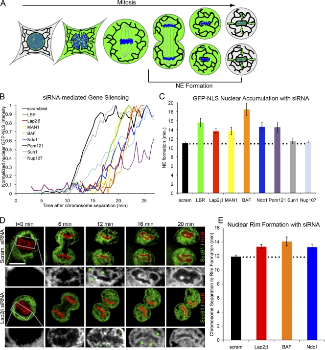
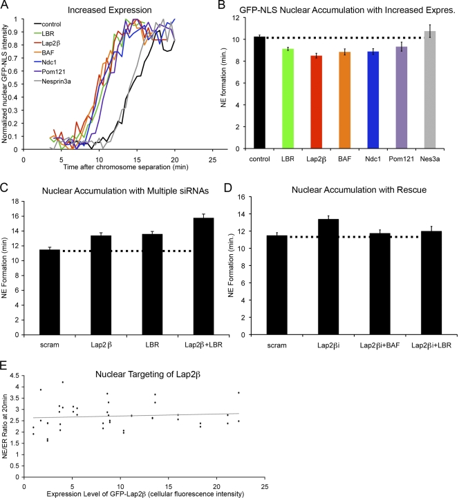
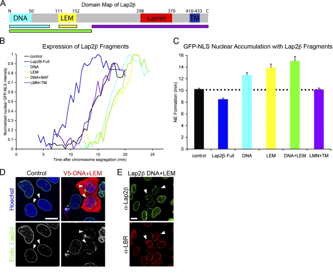
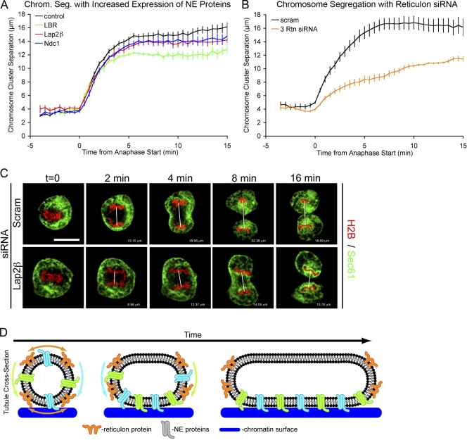
Formation of the nuclear envelope (NE) around segregated chromosomes occurs by the reshaping of the endoplasmic reticulum (ER), a reservoir for disassembled nuclear membrane components during mitosis. In this study, we show that inner nuclear membrane proteins such as lamin B receptor (LBR), MAN1, Lap2beta, and the trans-membrane nucleoporins Ndc1 and POM121 drive the spreading of ER membranes into the emerging NE via their capacity to bind chromatin in a collaborative manner. Despite their redundant functions, decreasing the levels of any of these trans-membrane proteins by RNAi-mediated knockdown delayed NE formation, whereas increasing the levels of any of them had the opposite effect. Furthermore, acceleration of NE formation interferes with chromosome separation during mitosis, indicating that the time frame over which chromatin becomes membrane enclosed is physiologically relevant and regulated. These data suggest that functionally distinct classes of chromatin-interacting membrane proteins, which are present at nonsaturating levels, collaborate to rapidly reestablish the nuclear compartment at the end of mitosis.


Figures

Figure 1.
Figure 1.
Chromatin-binding NE proteins collaborate during NE formation. (A) Diagram shows the dynamic localization of nuclear-targeted GFP (green) during open mitosis. Reaccumulation of GFP-NLS into daughter nuclei serves as an indicator for completed NE formation. (B) Cells were transfected with H2B-tdTomato and GFP-NLS and imaged through mitosis. Representative traces of chromatin-localized GFP-NLS in which t = 0 is set at the onset of chromosome separation show the time required for NE formation in U2OS cells with reduction of protein levels by siRNA knockdown. (C) Average time from chromosome separation to GFP-NLS nuclear accumulation was plotted. n > 20 for each condition (Table S1) with P < 0.01 when LBR, Lap2β, MAN1, BAF, Ndc1, or Pom121 siRNA was compared with scrambled (scram) RNA control, and P = 0.23 and 0.20 for Sun1 and Nup107, respectively (by t test). (D) U2OS cells were transfected with H2B-tdTomato (red) and Sec61-GFP (green, black, and white insets) and imaged from mitosis. Nuclear rim formation was compared in cells transfected with scrambled RNA or siRNA against Lap2β (closed arrowheads). After 12 min, no nuclear rim was detected with the knockdown of Lap2β (open arrowheads) compared with rim signal present in scrambled siRNA controls. Outlined areas represent the regions that are magnified below. Bar, 20 µm. (E) Average time from chromosome separation to complete nuclear rim formation was plotted. P < 0.01 when Lap2β, BAF, or Ndc1 knockdown was compared with scrambled RNA. Dotted lines indicate control cell timing. Error bars indicate SEM.
Figure 2.
Figure 2.
Chromatin-interacting NE proteins promote nuclear assembly. (A) Cells were transfected as in Fig. 1 B and imaged through mitosis. Representative traces of chromatin-localized GFP-NLS in which t = 0 is set at the onset of chromosome separation show the time required for NE formation in U2OS cells in which protein levels were increased by transfection with epitope-tagged constructs. (B) Average time from chromosome separation to GFP-NLS nuclear accumulation was plotted. P < 0.001 when Lap2β, LBR, BAF, Ndc1, or Pom121 increased expression (expres) was compared with control cells, and P = 0.20 for nesprin-3a (Nes3a; Table S2). (C) NE formation time was measured after partial knockdown of Lap2β, LBR, or both with a single round of siRNA transfection when Lap2β or LBR were compared with scrambled (scram) RNA oligos or when Lap2β + LBR was compared with Lap2β or LBR alone (P < 0.001; Table S3). (D) NE formation time was measured after partial knockdown of Lap2β combined with overexpression of either BAF or LBR and compared with the partial knockdown alone (P > 0.20 for each). (E) U2OS cells were transfected with GFP-Lap2β and H2B-tdTomato and imaged through mitosis. Average GFP fluorescence intensity was measured over entire cell and plotted against the ratio of GFP-Lap2β at the NE to peripheral GFP-Lap2β (NE/ER ratio). n > 20 for each condition (Table S3). Dotted lines indicate control cell timing. Error bars indicate SEM.
Figure 3.
Figure 3.
Membrane–chromatin tethering function of Lap2β in NE formation. (A) Map of Lap2β shows distinct functional domains that interact with DNA, BAF (LEM), lamins, and lipid bilayer (TM). (B) Representative traces of chromatin-localized GFP-NLS in which t = 0 is set at the onset of chromosome separation show the time required for NE formation in U2OS cells where fragments of Lap2β, DNA, LEM, DNA + LEM, or LMN + lipid bilayer have been overexpressed. (C) NE formation time was measured with the expression of Lap2β fragments. n > 40 for each fragment. P < 0.001 for the expression of DNA, LEM, and DNA + LEM fragments when compared with control cells; P = 0.4 for LMN + TM. Dotted line indicates control cell timing. Error bars indicate SEM. (D) U2OS cells were transfected with the V5-DNA + LEM fragment of Lap2β and stained with antibodies against V5 (red) and endogenous (endo) Lap2β (green). Arrowheads indicate early G1 cells as indicated by nuclear size and paired orientation. (E) U2OS cells were transfected with the DNA + LEM fragment of Lap2β and stained with antibodies against endogenous Lap2β and LBR. Arrowheads indicate cells where endogenous Lap2β, but not LBR, is displaced by the chromatin-binding domain of Lap2β. Bars, 20 µm.
Figure 4.
Figure 4.
Acceleration of nuclear membrane formation causes chromosome segregation defect. (A) Mitosis was analyzed by transfecting U2OS cells with Sec61-GFP and H2B-tdTomato and comparing control cells with cells in which NE formation was accelerated by overexpression of LBR, Lap2β, or Ndc1. Chromosome cluster separation (chrom seg) is plotted over time with P < 0.001 for Boltzmann Sigmoidal curve fitting to control cells. (B) Chromosome cluster separation plotted over time for extreme NE formation acceleration caused by the siRNA knockdown of reticulons 1, 3, and 4. (C) Representative images of U2OS cells with Sec61-GFP (green) and H2B-tdTomato (red) compares control cells with cells in which NE formation was accelerated by overexpression of Lap2β, and the distance between chromosome clusters was measured. t = 0 is set at anaphase onset. White lines indicate distances measured in Photoshop extended. Bar, 20 µm. (D) Cross-sectional schematic of a membrane tubule expanding onto chromatin (blue). Reticulons (orange) are displaced from the flat membrane where INM proteins (green) are targeted to chromatin and drive membrane expansion around chromatin. Error bars indicate SEM.

Similar articles

Cited by

References

    1. Anderson D.J., Hetzer M.W. 2007. Nuclear envelope formation by chromatin-mediated reorganization of the endoplasmic reticulum.Nat. Cell Biol. 9:1160–1166 - PubMed
    1. Anderson D.J., Hetzer M.W. 2008a. Reshaping of the endoplasmic reticulum limits the rate for nuclear envelope formation.J. Cell Biol. 182:911–924 - PMC - PubMed
    1. Anderson D.J., Hetzer M.W. 2008b. Shaping the endoplasmic reticulum into the nuclear envelope.J. Cell Sci. 121:137–142 - PubMed
    1. Antonin W., Ellenberg J., Dultz E. 2008. Nuclear pore complex assembly through the cell cycle: regulation and membrane organization.FEBS Lett. 582:2004–2016 - PubMed
    1. Burke B., Ellenberg J. 2002. Remodelling the walls of the nucleus.Nat. Rev. Mol. Cell Biol. 3:487–497 - PubMed

Publication types

MeSH terms