. 2009 Jul 27;186(2):173-82.
doi: 10.1083/jcb.200903100. Epub 2009 Jul 20.

The CENP-S complex is essential for the stable assembly of outer kinetochore structure

Affiliations

The CENP-S complex is essential for the stable assembly of outer kinetochore structure

Miho Amano et al. J Cell Biol. .

Abstract

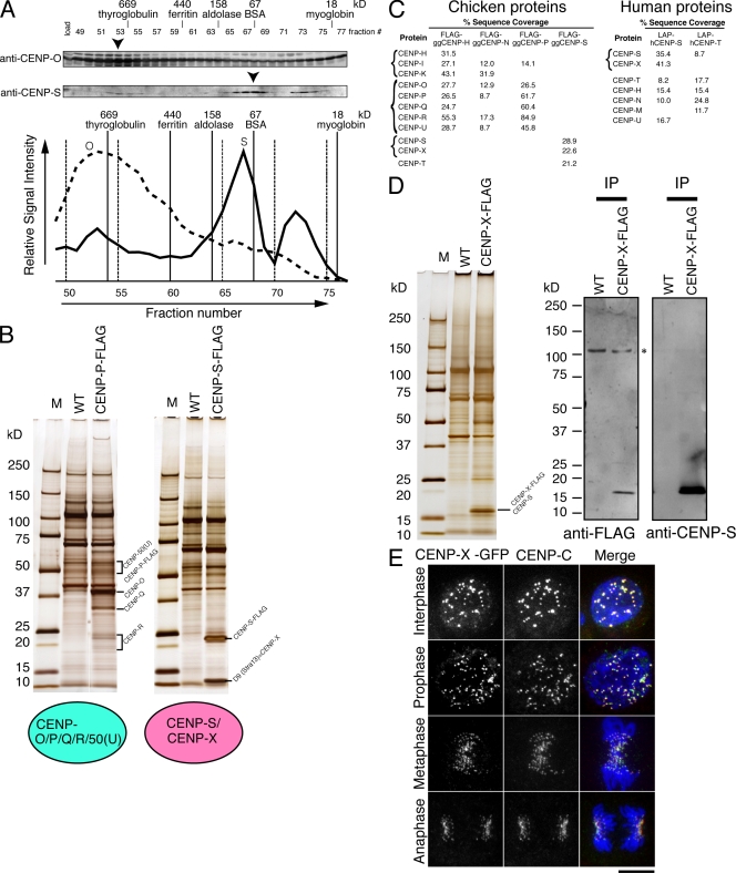
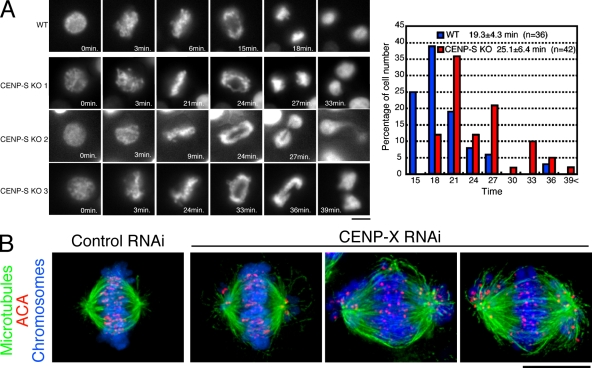
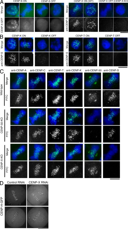
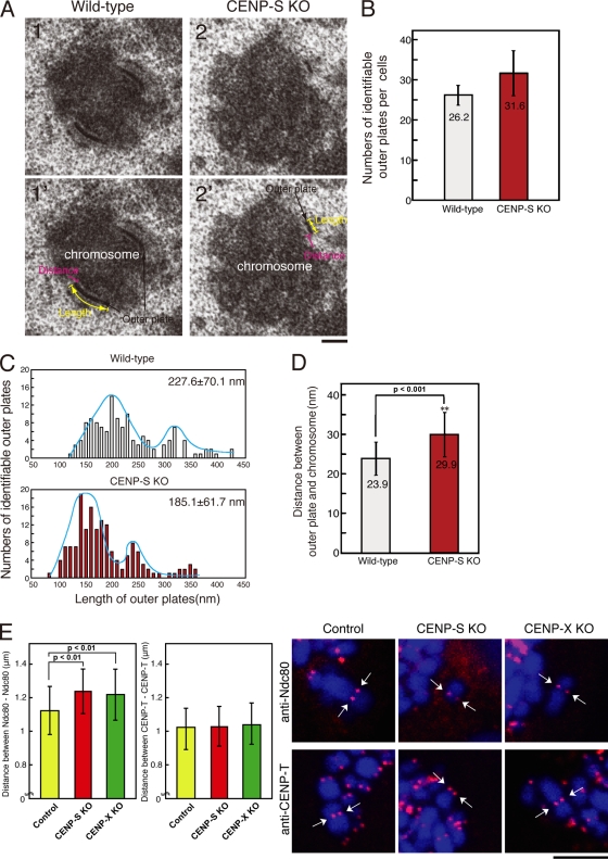
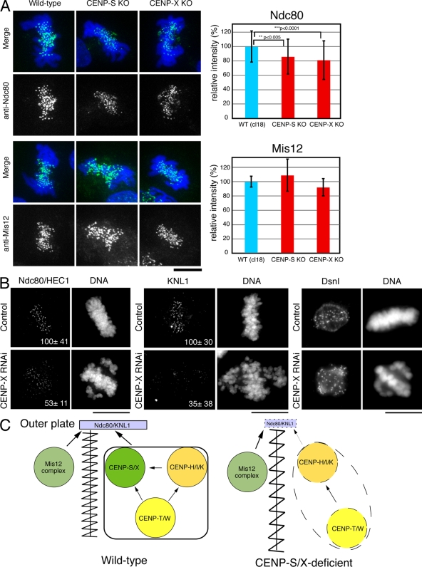
The constitutive centromere-associated network (CCAN) proteins are central to kinetochore assembly. To define the molecular architecture of this critical kinetochore network, we sought to determine the full complement of CCAN components and to define their relationships. This work identified a centromere protein S (CENP-S)-containing subcomplex that includes the new constitutive kinetochore protein CENP-X. Both CENP-S- and CENP-X-deficient chicken DT40 cells are viable but show abnormal mitotic behavior based on live cell analysis. Human HeLa cells depleted for CENP-X also showed mitotic errors. The kinetochore localization of CENP-S and -X is abolished in CENP-T- or CENP-K-deficient cells, but reciprocal experiments using CENP-S-deficient cells did not reveal defects in the localization of CCAN components. However, CENP-S- and CENP-X-deficient cells show a significant reduction in the size of the kinetochore outer plate. In addition, we found that intrakinetochore distance was increased in CENP-S- and CENP-X-deficient cells. These results suggest that the CENP-S complex is essential for the stable assembly of the outer kinetochore.


Figures

Figure 1.
Figure 1.
Identification of the CENP-S–associated protein CENP-X. (A) Immunoblots of DT40 cell extracts fractionated on a Superose 6 gel filtration column using antibodies against anti–CENP-O or –CENP-S. Signal intensities are plotted in the graph, and peak fractions are indicated by arrowheads. (B) SDS-PAGE of proteins isolated by IP with anti-Flag antibodies using cells in which expression of CENP-P or -S was replaced with CENP-P–Flag or CENP-S–Flag, respectively. Wild-type (WT) DT40 cells were also used for IP with anti-Flag antibodies as a control. (C) High sensitivity mass spectrometric analysis of the purifications of chicken and human centromere proteins indicating the percentage sequence coverage for each polypeptide. (D) Co-IP of CENP-S with CENP-X. Immunoprecipitates of CENP-X–expressing and wild-type cells with anti-Flag antibodies were separated by SDS-PAGE and analyzed by Western blotting with anti-Flag or –CENP-S antibodies. (E) Localization of GFP-tagged CENP-X throughout the cell cycle in DT40 cells. Centromeres were costained with anti–CENP-C antibodies. M, molecular mass marker. Bar, 10 µm.
Figure 2.
Figure 2.
Both CENP-S– and CENP-X–deficient DT40 cells are viable but show defects in mitotic progression. (A, left) Dynamics of chromosomes in wild-type (WT) or CENP-S–deficient cells visualized by time-lapse observation of living cells. Selected images of chromosomes from prophase to anaphase in these cells are shown. (right) Quantification of the time for progression from prophase to anaphase in wild-type and CENP-S–deficient cells as determined by time-lapse microscopy of living cells. (B) Chromosome morphology and α-tubulin staining (green) in human HeLa cells after siRNA-based knockdown for CENP-X. Human anticentromere antibodies (ACA) were used to detect the position of centromeres (red), and DNA (blue) was stained with Hoechst. Bars, 10 µm.
Figure 3.
Figure 3.
Kinetochore localization of the CENP-S–CENP-X complex occurs downstream of the CENP-H complex but is distinct from the CENP-O complex. (A) CENP-X localization in CENP-S–deficient cells (CENP-S OFF) and CENP-S localization in CENP-X–deficient cells (CENP-X OFF). WT, wild type. (B) CENP-S localization in CENP-K– or CENP-T–deficient cells (CENP-K OFF or CENP-T OFF, respectively). (C) Immunofluorescence analysis in wild-type DT40 and CENP-50– and CENP-X–deficient cells with the indicated antibodies (green). DNA was counterstained with DAPI (blue). (D) Imaging of human HeLa cells stably expressing GFP-tagged CENP-H after siRNA-based knockdown for control or CENP-X. Bars, 10 µm.
Figure 4.
Figure 4.
The length of the outer plate and the distance of the interkinetochore in CENP-S–deficient cells. (A) Electron micrograph showing a typical image of the kinetochore outer plate in wild-type (1 and 1′) or CENP-S–deficient cells (2 and 2′). (B) Numbers of identifiable outer plates per cell (mean ± SD). Approximately 35 serial sections were made for each cell, and the numbers of outer plates were counted. (C) Distribution of the outer plate length in wild-type or CENP-S–deficient cells. In both cell lines, two peaks with distribution were observed. The two peaks in the distribution of the plate length are likely caused by differences in sample orientation. As the orientation of some cells is not plane of the section, the length of plates varies depending on the angle of the section against a chromosome. (D) Distance between outer plate and electron-dense chromatin region in wild-type or CENP-S–deficient cells. (E) Distances between Ndc80 and Ndc80 or CENP-T and CENP-T of control or CENP-S– or CENP-X–deficient metaphase cells in the absence of microtubules. Arrows indicate sister kinetochore pairs. (D and E) Error bars represent SD. Bars: (A) 200 nm; (E) 5 µm.
Figure 5.
Figure 5.
Localization of outer plate proteins in CENP-S– or CENP-X–deficient cells. (A) Immunofluorescence analysis in wild-type (WT) DT40 or CENP-S– or CENP-X–deficient cells with anti-Ndc80 and -Mis12 antibodies. Signal intensities at each kinetochore were measured in these cells. Error bars represent SD. (B) Immunofluorescence analysis with anti-Ndc80, -KNL1, or -Dsn1 antibodies in HeLa cells treated with control or CENP-X siRNAs. The numbers in the micrographs are relative signal intensities of kinetochore signals. (C) A model for the assembly of kinetochore proteins at mitosis. The CENP-S–CENP-X complex is essential for the stable assembly of outer kinetochore proteins. In addition, the CENP-S–CENP-X complex generates a discrete CCAN structure to prevent the kinetochore from overstretching (left). In CENP-S– and CENP-X–deficient cells, the amount of KNL1 and Ndc80 at kinetochores is reduced, and CCAN structure is less tight, which causes kinetochore to be overstretched (right). Bars, 10 µm.

Similar articles

Cited by

References

    1. Cheeseman I.M., Desai A.. 2008. Molecular architecture of the kinetochore-microtubule interface.Nat. Rev. Mol. Cell Biol. 9:33–46. - PubMed
    1. Cheeseman I.M., Chappie J.S., Wilson-Kubalek E.M., Desai A.. 2006. The conserved KMN network constitutes the core microtubule-binding site of the kinetochore.Cell. 127:983–997. - PubMed
    1. Cheeseman I.M., Hori T., Fukagawa T., Desai A.. 2008. KNL1 and CENP-H/I/K complex coordinately direct kinetochore assembly in vertebrates.Mol. Biol. Cell. 19:587–594. - PMC - PubMed
    1. DeLuca J.G., Dong Y., Hergert P., Strauss J., Hickey J.M., Salmon E.D., McEwen B.F.. 2005. Hec1 and nuf2 are core components of the kinetochore outer plate essential for organizing microtubule attachment sites.Mol. Biol. Cell. 16:519–531. - PMC - PubMed
    1. DeLuca J.G., Gall W.E., Ciferri C., Cimini D., Musacchio A., Salmon E.D.. 2006. Kinetochore microtubule dynamics and attachment stability are regulated by Hec1.Cell. 127:969–982. - PubMed

Publication types

MeSH terms

Substances