. 2009 Jul 27;186(2):283-95.
doi: 10.1083/jcb.200905083. Epub 2009 Jul 20.

An outer arm dynein light chain acts in a conformational switch for flagellar motility

Affiliations

An outer arm dynein light chain acts in a conformational switch for flagellar motility

Ramila S Patel-King et al. J Cell Biol. .

Abstract

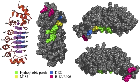
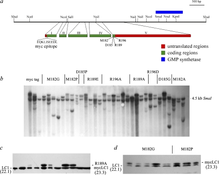
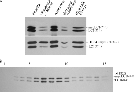
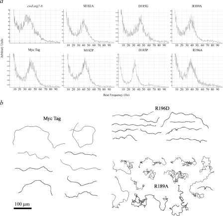
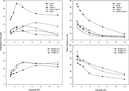
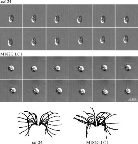
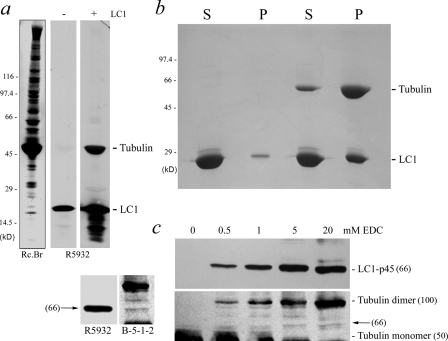
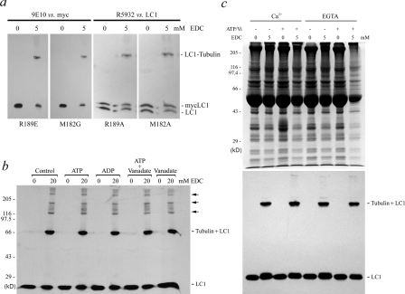
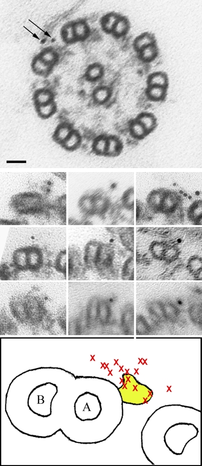
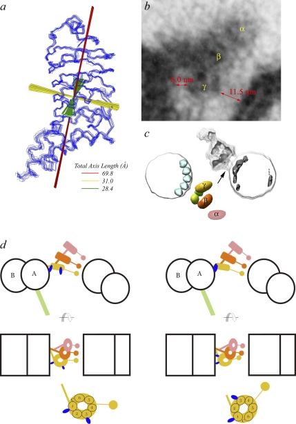
A system distinct from the central pair-radial spoke complex was proposed to control outer arm dynein function in response to alterations in the mechanical state of the flagellum. In this study, we examine the role of a Chlamydomonas reinhardtii outer arm dynein light chain that associates with the motor domain of the gamma heavy chain (HC). We demonstrate that expression of mutant forms of LC1 yield dominant-negative effects on swimming velocity, as the flagella continually beat out of phase and stall near or at the power/recovery stroke switchpoint. Furthermore, we observed that LC1 interacts directly with tubulin in a nucleotide-independent manner and tethers this motor unit to the A-tubule of the outer doublet microtubules within the axoneme. Therefore, this dynein HC is attached to the same microtubule by two sites: via both the N-terminal region and the motor domain. We propose that this gamma HC-LC1-microtubule ternary complex functions as a conformational switch to control outer arm activity.


Figures

Figure 1.
Figure 1.
Structure-based design of LC1 Mutants. The mean LC1 ribbon structure (Protein Data Bank accession no. 1M9L; left) and three views of the van der Waals molecular surface (right) are shown. The LRR region forms two β sheets and a helical face. The larger β sheet face (left, magenta) contains a single hydrophobic patch (green) centered on W99 that is predicted to bind the γ HC. The C-terminal portion of LC1 consists of a helical region, the orientation of which is controlled by two residues (M182 [yellow] and D185 [blue]) that show high backbone dynamics. The terminal α9 helix likely protrudes into the AAA+ domain and contains two Arg residues (R189 and R196; pink) that potentially make ionic contacts with the motor domain and/or nucleotide.
Figure 2.
Figure 2.
Expression of tagged mutant versions of LC1. (a) Map of the ∼6.2-kb LC1 genomic region, indicating the location of the five exons and the sites of myc tag insertion and mutagenesis. The genomic fragment also includes the gene for GMP synthase. (b) Southern blot analysis of SmaI-digested genomic DNA from strains transformed with various mutant forms of LC1. The endogenous LC1 gene yields a SmaI fragment of ∼4.5 kb (Benashski et al., 1999). The additional bands represent ≥1 integrated copies of the tagged LC1 gene. (c) Immunoblot analysis of flagellar samples from 11 strains transformed with the R189A mutant LC1 gene. The myc-tagged LC1 protein is detectable in five strains. (d) Immunoblot analysis of selected M182G- and M182P-transformed strains. These strains were chosen for analysis, as they stably express approximately similar amounts of wild-type and mutant LC1 proteins.
Figure 3.
Figure 3.
Myc-tagged LC1 copurifies with the outer dynein arm. (a) Immunoblots of flagellar fractions from strains expressing either myc-tagged LC1 or the D185G mutant form are shown. Addition of the myc tag alone (top) and most mutant forms did not alter either the detergent or 0.6 M NaCl extraction properties of LC1. In contrast, the D185G and M182G (not depicted) versions of LC1 exhibited an enhanced tendency to dissociate after detergent treatment of flagella (bottom). (b) Immunoblot of dynein from a strain expressing the M182G myc-tagged form of LC1 fractionated in a 5–20% sucrose density gradient; the bottom of the gradient is at left. The altered version of LC1 precisely comigrates with the native protein. Numbers in parentheses are given in kD.
Figure 4.
Figure 4.
Motile behavior of transformed strains. (a) The beat frequency power spectra for the cwd arg7-8 parental strain and transformants expressing myc-tagged LC1 and the M182A, M182P, D185G, D185P, R189A, and R196A mutant forms are shown. Strains incorporating the myc tag alone or the M182A, M182P, and D185G mutant forms have a beat frequency of ∼40–45, Hz which is similar to the parental strain. Both R189A and R196A mutants had beat frequencies reduced somewhat to 35–40 Hz, and only D185P exhibited a more serious reduction to ∼32 Hz. (b) The paths taken by individual cells expressing myc-tagged LC1 and the R196D and R189A mutant forms were tracked using MetaMorph. Most mutant strains moved in a helical path with occasional alterations in swimming direction as do wild-type cells. However, several R189A transformants swam very slowly and exhibited highly convoluted tracks as they continually altered direction.
Figure 5.
Figure 5.
Mutation of LC1 alters flagellar propulsive force generated under varying viscous load. (top) The beat frequency and propulsive force generated under varying viscous load is shown for wild type (cc124) and for strains lacking the entire outer arm (oda6), the motor domains of the β and γ HCs (oda4-s7 and oda2-t), and for a strain lacking both the β HC motor unit and the entire α HC (oda4-s7 oda11). (bottom) Similar plots for three strains expressing M182G, R189E, and R196A mutant forms of LC1 are shown. All three mutants generated reduced propulsive force under low viscous load, but this was enhanced as the load increased. This response approximates the behavior of the oda2-t mutant. Each point represents the mean propulsive force calculated from the swimming velocity of 10 cells. Bars indicating the SDs have been omitted for clarity; for all but two points on the cc124 and M182G plots, SDs ranged from 0.45–2.94 pN. Beat frequency data were obtained using the population-based fast Fourier transform method, and each determination was performed in triplicate.
Figure 6.
Figure 6.
Strains expressing mutant LC1 proteins exhibit alterations in flagellar phasing and the power/recovery stroke transition. Montages of individual sequential frames (1.67 ms apart) from high speed recordings illustrating the waveform of the cc124 wild type and a strain expressing the M182G mutant form of LC1 are shown. Flagellar of the mutant strain continually beat completely out of phase, leading to a rolling motion of the cell body of ∼50° in the plane of the flagella during the beat cycle. (bottom) Diagrams illustrating the superimposed waveforms are shown. Flagella containing mutant LC1 essentially stall for several milliseconds toward the end of the effective stroke, suggesting that they are inefficient at initiating the recovery stroke.
Figure 7.
Figure 7.
Association of LC1 and tubulin within the axoneme. (a) Nitrocellulose blots of axonemal proteins separated in a 5–15% acrylamide gradient gel were incubated in the presence or absence of recombinant LC1 and probed with the R5932 antibody. In the presence of LC1, the tubulin band was recognized, suggesting that LC1 binds tubulin. (left) The blot stained with Reactive brown 10 to detect total protein is shown. (b) Recombinant LC1 was incubated in the presence or absence of taxol-stabilized microtubules and spun in an airfuge for 5 min. Samples were electrophoresed in a 10% acrylamide gel and stained with Coomassie blue. In the absence of microtubules, only a very small fraction of LC1 was found in the pellet. However, upon microtubule addition, considerable LC1 bound microtubules and was present in the pellet fraction. Densitometry based on Coomassie dye binding indicated that the pellet contained a tubulin monomer/LC1 ratio of 1.00:1.06. (c) Axonemes were treated with 0–20 mM EDC, separated in an 8% acrylamide gel, blotted to nitrocellulose, and probed with R5932 and B-5-1-2 antibodies to detect LC1 and α-tubulin, respectively; note that most of the tubulin monomer band was excised from the blot before immunoblotting, as it produces a massive signal that otherwise obscures minor bands. The B-5-1-2 antibody detected two minor tubulin-containing products migrating between the tubulin monomer and dimer bands. The lower of these products precisely comigrated with the LC1-p45 band and was generated with similar kinetics. Numbers in parentheses indicate the approximate molecular mass of the indicated bands (given in kD).
Figure 8.
Figure 8.
Properties of LC1–tubulin interactions in situ. (a) Axonemes from strains expressing mutant myc-tagged forms of LC1 were treated with 0 or 5 mM EDC electrophoresed and immunoblotted using the 9E10 monoclonal antibody against the myc epitope or R5932 to detect LC1. In all strains examined, myc-tagged LC1 was readily incorporated into the Mr 66,000 LC1–tubulin cross-linked product, indicating that these proteins remain in direct contact. Furthermore, there were no obvious differences in the rate of incorporation of the tagged mutant forms compared with wild type, as both LC1 and myc-LC1 bands were reduced with similar kinetics. (b) Axonemes were treated with either 0 or 20 mM EDC in buffer or in the presence of 1 mM ATP, 1 mM ADP, 1 mM ATP plus 100 µM vanadate, and 100 µM vanadate alone. Samples were electrophoresed in a 5–15% acrylamide gradient gel, blotted to nitrocellulose, and probed with the R5932 antibody. The Mr 66,000 LC1–tubulin cross-linked product was generated by EDC treatment in similar yield under all nucleotide conditions, suggesting that LC1 and tubulin are permanently tethered to each other during the mechanochemical cycle. Several of the higher order products (arrows) show mass increase in steps of ∼50 kD and likely derive from cross-linking of the LC1–tubulin product to ≥1 additional tubulin molecules. (c) To test whether LC1–tubulin interactions were modified in a Ca2+-dependent manner, axonemes were resuspended in buffer containing 1 mM Ca2+ or EGTA in the presence or absence of 1 mM ATP/100 µM vanadate and were subsequently treated with 0 or 5 mM EDC. Samples were electrophoresed in a 10% acrylamide gel and stained with Coomassie blue (top) or probed with the R5932 antibody against LC1 (bottom). No differences in the yield of the LC1–tubulin product were observed as a consequence of Ca2+ addition.
Figure 9.
Figure 9.
Immunogold localization of LC1 within oda4-s7 oda11 axonemes. Electron micrographs of cross sections through an oda4-s7 oda11 axoneme (top) and individual outer doublets (middle) that had been incubated with antibody against LC1 and a secondary antibody conjugated to 5 nm gold. (bottom) A diagram illustrating the outer arm from this strain (yellow) and the location of individual gold particles (marked by x) is shown. The gold particles are clustered near the A-tubule of the doublet to which the outer arm is permanently attached. Arrows indicate two individual gold particles. Bar, 25 nm.
Figure 10.
Figure 10.
Geometry of the γ HC–LC1–microtubule ternary complex in situ. (a) The principal molecular axes of LC1 are superimposed on the ensemble of NMR-derived backbone structures (Protein Data Bank accession no. 1M9L) and the total axial dimensions indicated. LC1 is highly asymmetric with an axial ratio of 1.00 (green):1.13 (yellow):2.46 (red). (b) Thin section electron micrograph of the outer dynein arm in situ. The regions occupied by the motor domains of the three HCs are indicated as are the approximate distances between the γ HC motor unit and the neighboring outer doublet microtubules. (c) Three-dimensional reconstruction of the outer dynein arm–microtubule rigor complex from cryo-EM tomograms. Pink, α HC; orange, β HC; yellow, γ HC. There is a clear connection (arrow) between the γ HC motor unit and the microtubule to which the dynein is bound. The reconstruction is modified from Oda et al. (2007). (d) Models illustrating two potential geometries for the γ HC–LC1–microtubule complex in which LC1 is proposed to tether the γ HC to the A-tubule of the doublet to which dynein is permanently attached via the IC–LC complex and the docking complex. The HC color scheme is the same as in c. These models are based on the known dimensions of LC1, the measured motor unit/microtubule distances, and the immunogold localization of LC1 to the A-tubule. (left) The γ HC motor unit is in the same orientation as those of the α and β HCs. In this situation, it is unlikely that both copies of LC1 could interact simultaneously with the A-tubule. Thus, it is possible that the copy of LC1 that is bound switches as the AAA motor unit alters conformation during the mechanochemical cycle. (right) The γ HC motor unit is oriented differently such that both copies of LC1 bind the A-tubule simultaneously. In this situation, the system may act as a brake to limit sliding rather than as a microtubule translocase.

Similar articles

Cited by

References

    1. Baron D.M., Kabututu Z.P., Hill K.L. 2007. Stuck in reverse: loss of LC1 in Trypanosoma brucei disrupts outer dynein arms and leads to reverse flagellar beat and backward movement.J. Cell Sci. 120:1513–1520 - PubMed
    1. Bell C.W., Fronk E., Gibbons I.R. 1979. Polypeptide subunits of dynein 1 from sea urchin sperm flagella.J. Supramol. Struct. 11:311–317 - PubMed
    1. Belles-Isles M., Chapeau C., White D., Gagnon C. 1986. Isolation and characterization of dynein ATPase from bull spermatozoa.Biochem. J. 240:863–869 - PMC - PubMed
    1. Benashski S.E., Patel-King R.S., King S.M. 1999. Light chain 1 from the Chlamydomonas outer dynein arm is a leucine-rich repeat protein associated with the motor domain of the γ heavy chain.Biochemistry.38:7253–7264 - PubMed
    1. Bessen M., Fay R.B., Witman G.B. 1980. Calcium control of waveform in isolated flagellar axonemes of Chlamydomonas.J. Cell Biol. 86:446–455 - PMC - PubMed

Publication types

MeSH terms