. 2009 Jul 27;186(2):269-82.
doi: 10.1083/jcb.200901021. Epub 2009 Jul 20.

Membrane proteins follow multiple pathways to the basolateral cell surface in polarized epithelial cells

Affiliations

Membrane proteins follow multiple pathways to the basolateral cell surface in polarized epithelial cells

Glen A Farr et al. J Cell Biol. .

Abstract

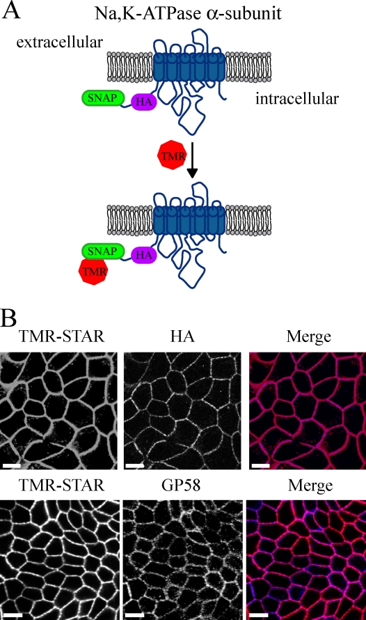
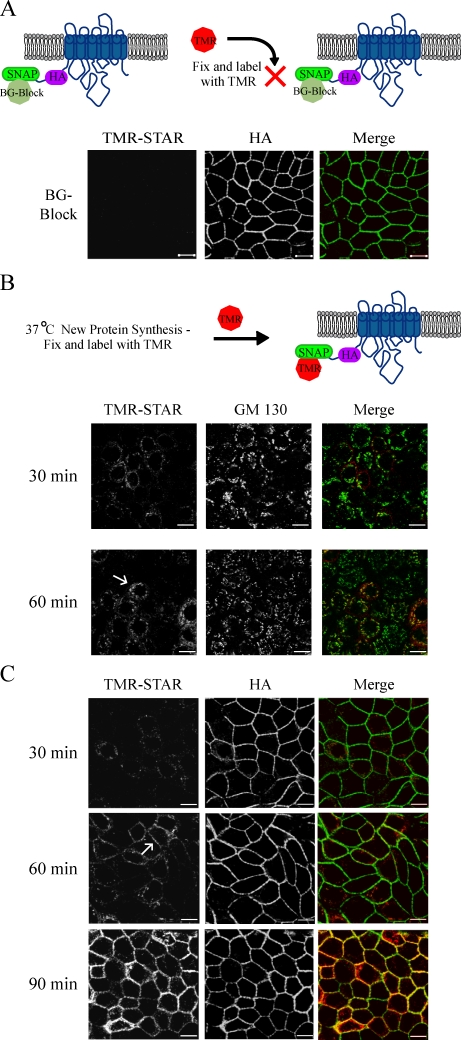
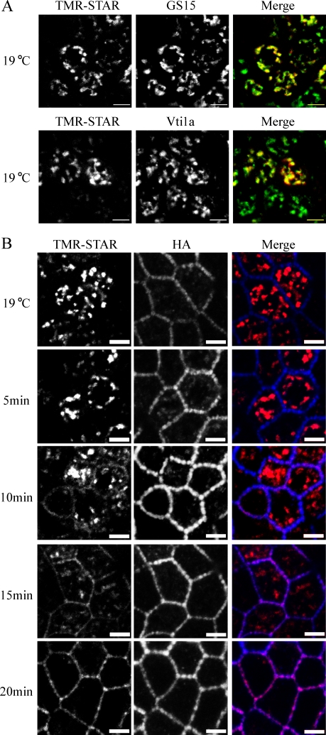
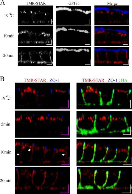
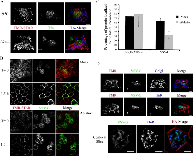
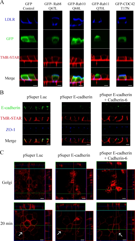
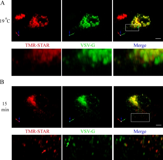
Newly synthesized apical and basolateral membrane proteins are sorted from one another in polarized epithelial cells. The trans-Golgi network participates in this sorting process, but some basolateral proteins travel from the Golgi to recycling endosomes (REs) before their surface delivery. Using a novel system for pulse-chase microscopy, we have visualized the postsynthetic route pursued by a newly synthesized cohort of Na,K-ATPase. We find that the basolateral delivery of newly synthesized Na,K-ATPase occurs via a pathway distinct from that pursued by the vesicular stomatitis virus G protein (VSV-G). Na,K-ATPase surface delivery occurs at a faster rate than that observed for VSV-G. The Na,K-ATPase does not pass through the RE compartment en route to the plasma membrane, and Na,K-ATPase trafficking is not regulated by the same small GTPases as other basolateral proteins. Finally, Na,K-ATPase and VSV-G travel in separate post-Golgi transport intermediates, demonstrating directly that multiple routes exist for transport from the Golgi to the basolateral membrane in polarized epithelial cells.


Figures

Figure 1.
Figure 1.
Detection of Na pump localization via the SNAP tag. (A) Diagram of the SNAP tag labeling reaction for the N-terminally tagged Na,K-ATPase α subunit. TMR, tetramethylrhodamine. (B) Stably transfected MDCK cells expressing both SNAP–Na,K-ATPase α subunit and Na,K-ATPase β subunit (SNAP cells) were fixed and stained with TMR-STAR (red) and processed for immunofluorescence with anti-HA or gp58 (blue). Bars, 10 µm.
Figure 2.
Figure 2.
Newly synthesized Na pump trafficking imaged via the SNAP tag. (A) Live SNAP cells were preincubated with nonfluorescent BG (BG-Block) for 30 min, fixed, stained with TMR-STAR (red), and processed for immunofluorescence with the indicated antibodies (green). (B and C) SNAP cells pretreated with BG-Block were washed and allowed to synthesize new protein for the indicated times at 37°C before fixation and labeling as in A. Arrows highlight examples of colocalization with the Golgi marker GM130 or with the lateral membrane stained with anti-HA. TMR, tetramethylrhodamine. Bars, 10 µm.
Figure 3.
Figure 3.
TGN-accumulated Na pump is rapidly trafficked to the lateral membrane. (A) SNAP cells were subjected to BG-Block, incubated at 37°C for 30 min to begin synthesis of new Na pump, and placed at 19°C for 2 h to accumulate newly synthesized protein in the TGN. TMR-STAR is depicted in red, and Golgi markers are shown in green. (B) SNAP cells were treated as in A and either fixed immediately (19°C) or warmed to 31°C for the indicated times. Samples were labeled with TMR-STAR (red) and with anti-HA (blue). Bars, 5 µm.
Figure 4.
Figure 4.
TGN-accumulated Na pump is trafficked directly to and randomly throughout the lateral membrane. SNAP cells were BG blocked, and newly synthesized Na,K-ATPase was accumulated in the TGN for 2 h at 19°C. After the Golgi block, samples were fixed immediately (19°C) or warmed to 31°C for the indicated times. (A) Samples labeled for TMR-STAR (red) and the apical membrane marker gp135 (blue). (B) Samples labeled with TMR-STAR (red), the junctional marker ZO-1 (blue), and for lateral membrane via anti-HA (green). Arrows denote areas of relatively high intensity TMR-STAR labeling along the lateral membrane. Bars, 5 µm.
Figure 5.
Figure 5.
The Na,K-ATPase does not pass through REs en route to the lateral membrane. (A) Cells were infected with adenovirus expressing the human Tfn-R, after which they were BG blocked, and newly synthesized Na pump was accumulated in the TGN (red) for 2 h at 19°C. Alexa Fluor 488–Tfn was added to the medium during the Golgi block to label REs (green). Golgi-blocked cells were fixed (19°C) or warmed for the indicated period and stained with anti-HA (blue). (B) Cells were infected to express the human Tfn-R and VSV-G–YFP Ts 045 (green) and incubated overnight at 40°C. Cells were BG blocked at 40°C and incubated for 40 min at 40°C with Tfn-HRP along with 1 µM TMR-STAR to live label newly synthesized pump (red). Next, cells were washed and incubated for a further 20 min at 40°C to remove excess TMR-STAR and to allow for the accumulation of Tfn-HRP into REs. Tfn-HRP–loaded REs were ablated on ice by exposing cells to DAB and H2O2 for 1 h, whereas control cells were treated with DAB alone. After the ablation reaction, cells were fixed or incubated in media plus CHX for 1.5 h at 31°C to allow trafficking of the proteins to the PM. (C) The percentage of protein that reached the lateral membrane 1.5 h after mock or ablation treatment was quantified using the enhanced colocalization tool (LSM Image Examiner software; Carl Zeiss, Inc.). 30 VSV-G–infected cells were analyzed, and data are represented as mean ± SD. (D) Samples were treated as in B and processed for immunofluorescence with a cocktail of Golgi antibodies (GM130, Vti1a, and GS15) or antibody against the Tfn-R shown in blue. The bottom panel depicts an individual confocal slice highlighting the colocalization of VSV-G with Tfn-R. TMR, tetramethylrhodamine. Bars, 5 µm.
Figure 6.
Figure 6.
Na pump trafficking is not regulated by the same small GTPases as AP-1B–dependent cargo and does not require E-cadherin. (A) SNAP cells were grown on clear polyester filters for 4 d to form a fully polarized monolayer and microinjected with plasmids expressing human LDLR and one of the following: GFP, GFP–Rab8 Q67L, GFP–Rab10 Q68L, GFP–Rab11 Q70L, or GFP–CDC42 T17N. Cells were then allowed to recover for 45 min before BG-Block addition. After BG blocking, cells were washed and allowed to synthesize new SNAP-tagged Na pump for 3 h, followed by incubation in the presence of CHX for 45 min. Samples were labeled with TMR-STAR (red) after being processed for surface immunofluorescence for LDLR (blue). (B). SNAP cells were transfected with shRNA expression plasmids directed against E-cadherin, Cadherin-6, or luciferase (Luc) as a negative control and cultured to allow monolayer polarization as previously described (Capaldo and Macara, 2007). 72 h after transfection, samples were stained with antibodies directed against E-cadherin (green) and ZO-1 (blue) and were labeled with TMR-STAR. (C) Cells were transfected and grown as in B and then subjected to the Golgi block protocol described in Fig. 3. Images depict samples fixed immediately after Golgi block or after incubation for 20 min at 31°C to allow Na pump exit from the TGN. Newly synthesized Na,K-ATPase traffics directly to the basolateral surface in cadherin-depleted cells. Arrows highlight lateral membrane delivery for each sample. Bars, 5 µm.
Figure 7.
Figure 7.
Na pump and VSV-G exit the TGN in separate PGTIs en route to the lateral membrane. SNAP cells were transfected with a plasmid encoding Ts 045 VSV-G–YFP and incubated for 24 h at 40°C to accumulate newly synthesized VSV-G in the ER. Next, samples were BG blocked at 40°C, washed, and incubated for a short 10-min recovery period to permit synthesis of new SNAP-tagged Na pump protein. (A and B) Samples were subjected to 19°C Golgi block and fixed immediately (19°C; A) or warmed to 31°C for 15 min (B). Samples were labeled with TMR-STAR (red) and imaged for YFP (green). The boxed areas are shown at higher magnification in the panels below. The green arrow highlights a PGTI containing only VSV-G, whereas the red arrow highlights a carrier containing only the Na pump. Bars, 5 µm.

Similar articles

Cited by

References

    1. Andreose J.S., Fumagalli G., Sigworth F.J., Caplan M.J. 1996. Real-time detection of the surface delivery of newly synthesized membrane proteins.Proc. Natl. Acad. Sci. USA. 93:7661–7666 - PMC - PubMed
    1. Ang A.L., Folsch H., Koivisto U.M., Pypaert M., Mellman I. 2003. The Rab8 GTPase selectively regulates AP-1B–dependent basolateral transport in polarized Madin-Darby canine kidney cells.J. Cell Biol. 163:339–350 - PMC - PubMed
    1. Ang A.L., Taguchi T., Francis S., Folsch H., Murrells L.J., Pypaert M., Warren G., Mellman I. 2004. Recycling endosomes can serve as intermediates during transport from the Golgi to the plasma membrane of MDCK cells.J. Cell Biol. 167:531–543 - PMC - PubMed
    1. Bok D., O'Day W., Rodriguez-Boulan E. 1992. Polarized budding of vesicular stomatitis and influenza virus from cultured human and bovine retinal pigment epithelium.Exp. Eye Res. 55:853–860 - PubMed
    1. Boll W., Partin J., Katz A., Caplan M., Jamieson J. 1991. Distinct pathways for basolateral targeting of membrane and secretory proteins in polarized epithelial cells.Proc. Natl. Acad. Sci. USA. 88:8592–8596 - PMC - PubMed

Publication types

MeSH terms