. 2009 Sep 25;284(39):26557-68.
doi: 10.1074/jbc.M109.025510. Epub 2009 Jul 20.

Structural and molecular mechanism for autoprocessing of MARTX toxin of Vibrio cholerae at multiple sites

Affiliations

Structural and molecular mechanism for autoprocessing of MARTX toxin of Vibrio cholerae at multiple sites

Katerina Prochazkova et al. J Biol Chem. .

Abstract

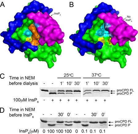
The multifunctional autoprocessing repeats-in-toxin (MARTX) toxin of Vibrio cholerae causes destruction of the actin cytoskeleton by covalent cross-linking of actin and inactivation of Rho GTPases. The effector domains responsible for these activities are here shown to be independent proteins released from the large toxin by autoproteolysis catalyzed by an embedded cysteine protease domain (CPD). The CPD is activated upon binding inositol hexakisphosphate (InsP(6)). In this study, we demonstrated that InsP(6) is not simply an allosteric cofactor, but rather binding of InsP(6) stabilized the CPD structure, facilitating formation of the enzyme-substrate complex. The 1.95-A crystal structure of this InsP(6)-bound unprocessed form of CPD was determined and revealed the scissile bond Leu(3428)-Ala(3429) captured in the catalytic site. Upon processing at this site, CPD was converted to a form with 500-fold reduced affinity for InsP(6), but was reactivated for high affinity binding of InsP(6) by cooperative binding of both a new substrate and InsP(6). Reactivation of CPD allowed cleavage of the MARTX toxin at other sites, specifically at leucine residues between the effector domains. Processed CPD also cleaved other proteins in trans, including the leucine-rich protein YopM, demonstrating that it is a promiscuous leucine-specific protease.


Figures

FIGURE 1.
FIGURE 1.
Structural model of pro-CPD/C-S reveals enzyme-substrate complex. A, pro-CPD/C-S with N terminus (blue), protease core (green), β-flap (magenta), and InsP6 (red). Key residues (orange) and catalytic residues (yellow) are labeled according to annotation of Lin et al. (24). B, schematic representation of the Clan CD fold catalytic site with P1 Leu3428 (magenta) inserted into S1 site. Distances (in angstroms) of key bonds are shown as dashed lines. C, stereo view of the active site of pro-CPD/C-S as a stick model with surrounding 2FoFc map contoured at 1 sigma (green) and the N terminus residues, surrounded with omit FoFc map contoured at 4 sigma level (blue; omitted residues are Ala-Leu-Ala). For B and C, carbon of the active site, carbon of the substrate, oxygen and nitrogen atoms are colored in green, yellow, red, and blue, respectively.
FIGURE 2.
FIGURE 2.
rCPD cleaves a Leu-Xaa bond. A, Coomassie-stained gel of autoprocessing of rCPD L3428A (FL) shows processing at indicated concentrations of InsP6 after 2-h incubation resulted in a shift of autocleavage site to generate a slightly longer fragment (marked HLQ) than the normal processing site (marked ALA). B, the size of the processed product (P) is not affected by alteration of P2 or P1′ Ala residues in full-length rCPD (FL). C, the predominant processing sites in various point mutants (double underlined) are shown by line bracket.
FIGURE 3.
FIGURE 3.
Binding of InsP6 increases pro-CPD/C-S Tm, and the protein becomes trypsin-resistant. A, close-up view of InsP6 binding pocket shows 12 residues known to contact InsP6 (red with space-filling dots) derive from the N terminus (blue), the protease core (green), and the β-flap (magenta). B, SYPRO® Orange melting curves of pro-CPD/C-S at different concentrations of InsP6. C, Coomassie-stained gel of limited proteolysis of pro-CPD/C-S at varying concentrations of trypsin. Locations of trypsin cleavage in the absence of InsP6 as determine by FT-MS (supplemental Fig. 4) are shown in orange in D with color scheme used in A except antiparallel β8β9 are highlighted pink and S1 hydrophobic residues are space-filling dots.
FIGURE 4.
FIGURE 4.
Susceptibility of pro-CPD to inhibition by NEM depends on position of the N terminus. A and B, space-filling models of pro-CPD/C-S with N terminus (blue), protease core (green), β-flap (magenta), and InsP6 (red). Models are based on the B molecule. In A, P1 Leu3428 (L) is buried in the hydrophobic S1 site (formed by the protease core (aqua) and the β-flap (medium blue)). The catalytic cysteine (CS) is covered by the N terminus at the scissile bond (dashed line) and by Glu3602 (E). In B, removal of the N terminus to the P3′ residue exposes the catalytic cysteine. Other labeled residues are the catalytic His3519 (H), S1 residues from the α1 helix (Val3472V) and Ala3475A)) or the β8 strand (Leu3603L) and Val3605V)) and the N terminus residues in the binding cleft identified by single letter code. C, pro-CPD with intact cysteine was incubated without (−) or with 100 μm NEM for time indicated at temperature indicated after which the protein was dialyzed at 4 °C to remove excess inhibitor. Processing was then initiated by addition of 100 μm InsP6 and processing of full-length protein (FL) to the processed form (P) was assessed by SDS-PAGE. D, pro-CPD was incubated without NEM (−) or with 100 μm NEM for 30 min (30′) at 25 °C after which InsP6 as indicated was added. In lanes marked 0′, NEM and InsP6 were first mixed and then added simultaneously (0 min preincubation). Reactions were monitored after 1 h at 37 °C by SDS-PAGE.
FIGURE 5.
FIGURE 5.
Post-CPD can be reactivated for InsP6 binding and trypsin resistance by CK inhibitors. A, purified post-CPD was treated with trypsin in the presence of the indicated concentration of InsP6 and proteolysis was determined by SDS-PAGE. The first cleavage band arising in the 0.1-μg lane corresponds to processing at Arg3610 or Lys3611, with the second band corresponding to processing at Arg3599. Preincubation of post-CPD with 100 μm l-LeuCK (L) or z-Gly-Leu-Phe-CK (GLF) protects the protein from trypsin while T-LysCK (K) is only partially protected. B, ITC curves performed with 15 μm post-CPD as described under “Experimental Procedures” either without or with preincubation of 100 μm CK inhibitors as noted. The pro-CPD/C-S control is shown in greater detail in supplemental Fig. 2.
FIGURE 6.
FIGURE 6.
Post-CPD can process rCPD/C-S and unrelated proteins in trans. A, Coomassie-stained gels of full-length mutant rCPD/C-S proteins (rCPD FL) that cannot autoprocess. If the S1 site (L3479D) or InsP6 binding pocket (K3611A and K3482A) are modified to prevent Leu from occupying its own active site, mutants can be processed (P) by post-CPD dependent upon InsP6. B, full-length proteins (FL) were incubated without inhibitor (−) or with 100 μm NEM (N) or l-LeuCK (L) for time specified at 25 °C after which proteins were dialyzed to remove excess inhibitor, and 100 μm InsP6 was added when indicated to initiate processing of rCPD L3479D/C3568S by post-CPD. Processing for both assays was determined by SDS-PAGE after 2-h incubation at 37 °C. For C purified YopM was incubated with pro-CPD in 0.4 m urea and with or without 100 μm InsP6 for the times indicated.
FIGURE 7.
FIGURE 7.
CPD domain autoprocesses RtxA1580–3909 in multiple sites. A and B, NEM-treated RtxA1580–3909 can serve as a substrate for CPD (lanes 1–6), but also untreated RtxA1580–3909 can undergo autoprocessing (lanes 5–7). For lanes 1–6, RtxA1580–3909 was first incubated with 1 mm NEM for 30 min at 25 °C, after which protein was dialyzed to remove excess of inhibitor. Then, pro-CPD was mixed with NEM-pretreated RtxA1580–3909 and incubated for 5 min, after which processing was initiated by addition of 100 μm InsP6 for the time indicated at 37 °C. The asterisk in lane 6 marks a fragment absent after autoprocessing (lane 9). For lanes 7–9 and B, autoprocessing reactions were initiated by addition of 100 μm InsP6 and terminated at the times indicated. In B, protein was incubated with 1 mm of the indicated inhibitor for 30 min at 25 °C prior to addition of InsP6. C and D, N termini of marked fragments (F1–F5) arising from autoprocessing for 1 min were identified by Edman degradation and are marked with the first five amino acids (C) and correspond to fragments diagrammed in D. E, actin cross-linking reactions were performed with commercial G-actin in the presence of InsP6 and/or 5 mm EDTA as indicated. The arrowhead marks RtxA1580–3909. F, alignment of all processing sites identified in this study.

Similar articles

Cited by

References

    1. Satchell K. J. (2007) Infect. Immun. 75, 5079–5084 - PMC - PubMed
    1. Olivier V., Haines G. K., 3rd, Tan Y., Satchell K. J. (2007) Infect. Immun. 75, 5035–5042 - PMC - PubMed
    1. Olivier V., Salzman N. H., Satchell K. J. (2007) Infect. Immun. 75, 5043–5051 - PMC - PubMed
    1. Kudryashov D. S., Durer Z. A., Ytterberg A. J., Sawaya M. R., Pashkov I., Prochazkova K., Yeates T. O., Loo R. R., Loo J. A., Satchell K. J., Reisler E. (2008) Proc. Natl. Acad. Sci. U.S.A. 105, 18537–18542 - PMC - PubMed
    1. Sheahan K. L., Cordero C. L., Satchell K. J. (2004) Proc. Natl. Acad. Sci. U.S.A. 101, 9798–9803 - PMC - PubMed

Publication types

MeSH terms

Associated data

LinkOut - more resources