. 2009 Aug 4;106(31):12682-7.
doi: 10.1073/pnas.0904843106. Epub 2009 Jul 20.

The JNKs differentially regulate RNA polymerase III transcription by coordinately modulating the expression of all TFIIIB subunits

Affiliations

The JNKs differentially regulate RNA polymerase III transcription by coordinately modulating the expression of all TFIIIB subunits

Shuping Zhong et al. Proc Natl Acad Sci U S A. .

Abstract

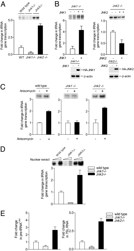
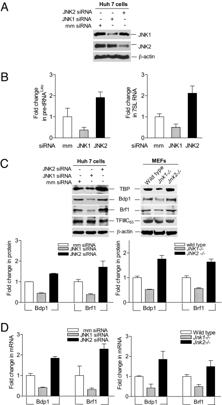
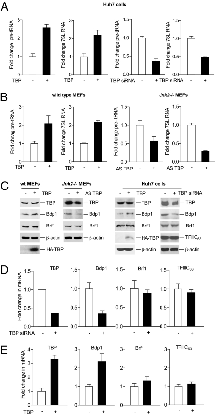
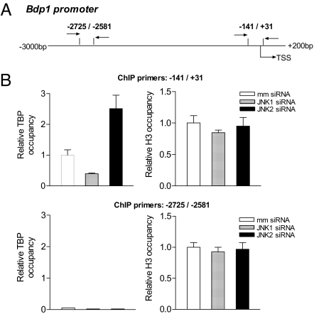
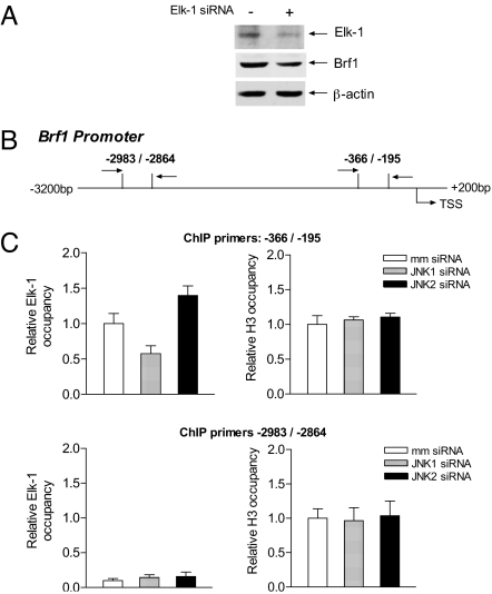
RNA polymerase (pol) III-dependent transcription is subject to stringent regulation by tumor suppressors and oncogenic proteins and enhanced RNA pol III transcription is essential for cellular transformation and tumorigenesis. Since the c-Jun N-terminal kinases (JNKs) display both oncogenic and tumor suppressor properties, the roles of these proteins in regulating RNA pol III transcription were examined. In both mouse and human cells, loss or reduction in JNK1 expression represses RNA pol III transcription. In contrast, loss or reduction in JNK2 expression induces transcription. The JNKs coordinately regulate expression of all 3 TFIIIB subunits. While JNK1 positively regulates TBP expression, the RNA pol III-specific factors, Brf1 and Bdp1, JNK2 negatively regulates their expression. Brf1 is coregulated with TBP through the JNK target, Elk-1. Reducing Elk-1 expression decreases Brf1 expression. Decreasing JNK1 expression reduces Elk-1 occupancy at the Brf1 promoter, while decreasing JNK2 expression enhances recruitment of Elk-1 to the Brf1 promoter. In contrast, regulation of Bdp1 occurs through JNK-mediated alterations in TBP expression. Altered TBP expression mimics the effect of reduced JNK1 or JNK2 levels on Bdp1 expression. Decreasing JNK1 expression reduces the occupancy of TBP at the Bdp1 promoter, while decreasing JNK2 expression enhances recruitment of TBP to the Bdp1 promoter. Together, these results provide a molecular mechanism for regulating RNA pol III transcription through the coordinate control of TFIIIB subunit expression and elucidate opposing functions for the JNKs in regulating a large class of genes that dictate the biosynthetic capacity of cells.


Conflict of interest statement

The authors declare no conflict of interest.

Figures

Fig. 1.
Fig. 1.
RNA pol III transcription is differentially regulated by JNK1 and JNK2 in MEFs. (A) RNA pol III transcription is altered in MEFs deficient for Jnk1 or Jnk2. Wild-type, Jnk1−/−, and Jnk2−/− MEFs were transiently transfected with a tRNA gene reporter plasmid. RNA was extracted, and RNase protection assays were carried out. Quantification of 3 independent experiments is shown, and an example of an autoradiogram is shown above the graph. (B) Expression of JNK1 increases, while JNK2 represses, transcription. MEFs were cotransfected with expression plasmids for HA-JNK1 or HA-JNK2 with the tRNA gene reporter. RNA analysis was performed as in A Upper, and immunoblot analysis was used to detect the HA-JNKs using HA antibodies (Lower). Membranes were stripped and reprobed with antibodies against β-actin. (C) Anisomycin induction of tRNA transcription requires JNK1. MEFs were transfected with a tRNA gene reporter and treated with or without anisomycin before isolation. tRNA gene transcription was measured as in A. (D) JNK-mediated regulation of tRNA gene transcription can be reproduced in vitro. Nuclear proteins were prepared from MEFs, and in vitro transcription reactions were carried out using the tRNA reporter template and 10 or 20 μg nuclear extract. (E) Endogenous RNA pol III transcription is altered in MEFs deficient for Jnk1 or Jnk2. RT-qPCR was performed using RNA isolated from wild-type, Jnk1−/−, and Jnk2−/− MEFs and with specific primers for precursor (pre) tRNALeu or 7SL RNA. Quantification of 3 independent experiments from 3 independent RNA isolations is shown.
Fig. 2.
Fig. 2.
Expression of all TFIIIB subunits are coordinately regulated by the JNKs. (A) siRNAs specifically repress JNK1 or JNK2 expression. Protein lysates derived from Huh-7 cells transfected with siRNAs against JNK1 or JNK2 were subjected to immunoblot analysis using antibodies against JNK1, JNK2, or β-actin as indicated. (B) RNA pol III transcription is differentially regulated by JNK1 or JNK2 in Huh-7 cells. Huh-7 cells were transfected with siRNAs for JNK1, JNK2, or mismatch RNA. RNA was isolated from the transfected cells, and real time RT-qPCR was performed using specific primers for precursor (pre) tRNALeu (Left) or 7SL RNA (Right). (C) JNK1 and JNK2 differentially regulate expression of TFIIIB subunits, TBP, Bdp1, and Brf1. Immunoblot analysis was preformed using lysates derived from either siRNA transfected Huh-7 cells (Upper Left) or from MEFs (Upper Right). Antibodies against TBP, Bdp1, Brf1, TFIIIC63, or β-actin were used as indicated. Quantification of protein amounts from 3 independent experiments relative to β-actin is shown Bdp1 and Brf1 for results obtained using Huh-7 (Lower Left) and MEFs (Lower Right). (D) JNK1 and JNK2 differentially regulate Bdp1 and Brf1 mRNAs. RT-qPCR was used to measure Bdp1 and Brf1 mRNAs using RNA derived from siRNA transfected Huh-7 cells or from MEFs as described in B.
Fig. 3.
Fig. 3.
Changes in the cellular level of TBP modulates Bdp1 expression and RNA pol III transcription. (A) Changes in TBP expression modulate transcription of tRNALeu and 7SL RNA in Huh-7 cells. Cells were transfected with a human TBP expression plasmid (Left) or a TBP siRNA (Right). Real-time RT-PCR was performed using isolated RNA and primers specific for pretRNALeu or 7SL RNA, and the fold change was calculated by normalizing to the amount of GAPDH mRNA. (B) Changes in TBP expression modulate transcription of tRNALeu and 7SL RNA in MEFs. Wild-type MEFs were transfected with TBP expression plasmid and amounts of pretRNALeu and 7SL RNA were measured (Left), and Jnk2−/− MEFs were transfected with antisense TBP construct and the amount of pretRNALeu and 7SL RNA was measured as in A. (C) Alterations in TBP expression changes Bdp1, but not Brf1 expression. Wild-type MEFs were transfected with a TBP expression plasmid and Jnk2−/− MEFs with an antisense TBP construct (Left). Huh-7 cells were transfected with either an siRNA against TBP or an expression plasmid for HA-tagged TBP (Right). Resulting lysates were subjected to immunoblot analysis using antibodies against TBP, Bdp1, Brf1, TFIIIC63, β-actin, or HA-TBP as designated. (D) Reduction in cellular TBP expression decreases Bdp1, but not Brf1, mRNA levels in Huh-7 cells. Cells were transfected with an siRNA against TBP, RNA was isolated, and RT-qPCR was conducted to measure the amounts of TBP, Bdp1, Brf1, and TFIIIC63 mRNA. Three independent experiments were performed, and the fold change was normalized to GAPDH mRNA. (E) Enhanced expression of TBP increases Bdp1, but not Brf1, mRNA levels in Huh-7 cells. Cells were transfected with a TBP expression plasmid, and RT-qPCR was performed to quantify the amounts of mRNAs for TBP, Bdp1, Brf1, and TFIIIC63. Three independent experiments were performed, and the fold change was normalized to GAPDH mRNA.
Fig. 4.
Fig. 4.
JNK1 and JNK2 differentially regulate TBP occupancy on the Bdp1 promoter. (A) Schematic of the Bdp1 promoter. The diagram depicts the start site for transcription on the human Bdp1 promoter (TSS) identified by using a RACE kit (Invitrogen). The 2 primer pairs were used to amplify the PCR products, 1 at (−142 bp/+31 bp) encompassing the TSS and an upstream fragment (−2725 bp/-2581 bp). (B) TBP occupancy on the Bdp1 promoter is regulated by the JNKs. Huh-7 cells were transfected with siRNAs for JNK1, JNK2, or mismatch siRNAs. ChIP assays were performed using antibodies against TBP or histone H3 and the primer pair -141/31 encompassing the transcription start site (Upper) or the primer pair -2725/-2581 upstream of the transcription start site. The bars represent ±SE of at least 3 independent determinations from 3 separate chromatin preparations. The occupancy of TBP or histone H3 was calculated relative to the percent occupancy observed in cells transfected with mismatch RNA.
Fig. 5.
Fig. 5.
JNK1 and JNK2 differentially regulate Elk-1 occupancy on the Brf1 promoter. (A) Decreased Elk-1 expression reduces Brf1 expression. Huh-7 cells were transfected with siRNAs specific for Elk-1 (+) or mismatch RNA (-), and immunoblot analysis was used to determine the amount of Elk-1, Brf1, and β-actin. (B) Schematic of the Brf1 promoter. The diagram depicts the start site for transcription on the human Brf1 promoter identified previously (22). The 2 primer pairs used to amplify the ChIP PCR products at -366/-195 and an upstream fragment at -2983/-2864 are depicted. (C) The JNKs have opposing affects on Elk-1 occupancy on the Brf1 promoter. Huh-7 cells were transfected with siRNAs for JNK1, JNK2, or mismatch siRNAs. ChIP assays were performed using antibodies against Elk-1 or histone H3 and the primer pair encompassing -366/-195 (top) or the -2983/-2864 (bottom) relative to the transcription start site. The bars represent ±SE of 4 independent determinations from 3 separate chromatin preparations. The occupancy of Elk-1 or histone H3 was calculated relative to the percent occupancy observed in cells transfected with mismatch RNA.

Similar articles

Cited by

References

    1. Chang L, Karin M. Mammalian MAP kinase signalling cascades. Nature. 2001;410:37–40. - PubMed
    1. Davis RJ. Signal transduction by the JNK group of MAP kinases. Cell. 2000;103:239–252. - PubMed
    1. Bode AM, Dong Z. The functional contrariety of JNK. Mol Carcinog. 2007;8:591–598. - PMC - PubMed
    1. Ventura JJ, Hübner A, Zhang C, Flavell RA, Shokat KM, Davis RJ. Chemical genetic analysis of the time course of signal transduction by JNK. Mol Cell. 2006;5:701–710. - PubMed
    1. Wang H-D, Trivedi A, Johnson DL. Hepatitis B virus X protein induces RNA polymerase III-dependent gene transcription and increases cellular TATA-binding protein by activating the Ras signaling pathway. Mol Cell Biol. 1997;17:6838–6846. - PMC - PubMed

Publication types

MeSH terms

Substances

LinkOut - more resources