The N-end rule pathway controls multiple functions during Arabidopsis shoot and leaf development
- PMID: 19620738
- PMCID: PMC2726413
- DOI: 10.1073/pnas.0906404106
The N-end rule pathway controls multiple functions during Arabidopsis shoot and leaf development
Abstract
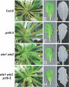
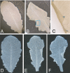
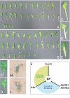
The ubiquitin-dependent N-end rule pathway relates the in vivo half-life of a protein to the identity of its N-terminal residue. This proteolytic system is present in all organisms examined and has been shown to have a multitude of functions in animals and fungi. In plants, however, the functional understanding of the N-end rule pathway is only beginning. The N-end rule has a hierarchic structure. Destabilizing activity of N-terminal Asp, Glu, and (oxidized) Cys requires their conjugation to Arg by an arginyl-tRNA-protein transferase (R-transferase). The resulting N-terminal Arg is recognized by the pathway's E3 ubiquitin ligases, called "N-recognins." Here, we show that the Arabidopsis R-transferases AtATE1 and AtATE2 regulate various aspects of leaf and shoot development. We also show that the previously identified N-recognin PROTEOLYSIS6 (PRT6) mediates these R-transferase-dependent activities. We further demonstrate that the arginylation branch of the N-end rule pathway plays a role in repressing the meristem-promoting BREVIPEDICELLUS (BP) gene in developing leaves. BP expression is known to be excluded from Arabidopsis leaves by the activities of the ASYMMETRIC LEAVES1 (AS1) transcription factor complex and the phytohormone auxin. Our results suggest that AtATE1 and AtATE2 act redundantly with AS1, but independently of auxin, in the control of leaf development.
Conflict of interest statement
The authors declare no conflict of interest.
Figures


Similar articles
-
Meta-analyses of microarrays of Arabidopsis asymmetric leaves1 (as1), as2 and their modifying mutants reveal a critical role for the ETT pathway in stabilization of adaxial-abaxial patterning and cell division during leaf development.Plant Cell Physiol. 2013 Mar;54(3):418-31. doi: 10.1093/pcp/pct027. Epub 2013 Feb 8. Plant Cell Physiol. 2013. PMID: 23396601 Free PMC article.
-
ASYMMETRIC LEAVES1 and auxin activities converge to repress BREVIPEDICELLUS expression and promote leaf development in Arabidopsis.Development. 2006 Oct;133(20):3955-61. doi: 10.1242/dev.02545. Epub 2006 Sep 13. Development. 2006. PMID: 16971475
-
The Arabidopsis BEL1-LIKE HOMEODOMAIN proteins SAW1 and SAW2 act redundantly to regulate KNOX expression spatially in leaf margins.Plant Cell. 2007 Sep;19(9):2719-35. doi: 10.1105/tpc.106.048769. Epub 2007 Sep 14. Plant Cell. 2007. PMID: 17873098 Free PMC article.
-
The complex of ASYMMETRIC LEAVES (AS) proteins plays a central role in antagonistic interactions of genes for leaf polarity specification in Arabidopsis.Wiley Interdiscip Rev Dev Biol. 2015 Nov-Dec;4(6):655-71. doi: 10.1002/wdev.196. Epub 2015 Jun 24. Wiley Interdiscip Rev Dev Biol. 2015. PMID: 26108442 Free PMC article. Review.
-
The shady side of leaf development: the role of the REVOLUTA/KANADI1 module in leaf patterning and auxin-mediated growth promotion.Curr Opin Plant Biol. 2017 Feb;35:111-116. doi: 10.1016/j.pbi.2016.11.016. Epub 2016 Dec 3. Curr Opin Plant Biol. 2017. PMID: 27918939 Review.
Cited by
-
Waterproofing crops: effective flooding survival strategies.Plant Physiol. 2012 Dec;160(4):1698-709. doi: 10.1104/pp.112.208173. Epub 2012 Oct 23. Plant Physiol. 2012. PMID: 23093359 Free PMC article. Review. No abstract available.
-
The N-end rule pathway and regulation by proteolysis.Protein Sci. 2011 Aug;20(8):1298-345. doi: 10.1002/pro.666. Protein Sci. 2011. PMID: 21633985 Free PMC article. Review.
-
A Shoot-Specific Hypoxic Response of Arabidopsis Sheds Light on the Role of the Phosphate-Responsive Transcription Factor PHOSPHATE STARVATION RESPONSE1.Plant Physiol. 2014 Jun;165(2):774-790. doi: 10.1104/pp.114.237990. Epub 2014 Apr 21. Plant Physiol. 2014. PMID: 24753539 Free PMC article.
-
The N-end rule and retroviral infection: no effect on integrase.Virol J. 2013 Jul 13;10:233. doi: 10.1186/1743-422X-10-233. Virol J. 2013. PMID: 23849394 Free PMC article.
-
Biochemical analysis of protein arginylation.Methods Enzymol. 2019;626:89-113. doi: 10.1016/bs.mie.2019.07.028. Epub 2019 Aug 1. Methods Enzymol. 2019. PMID: 31606094 Free PMC article.
References
-
- Bachmair A, Varshavsky A. The degradation signal in a short-lived protein. Cell. 1989;56:1019–1032. - PubMed
-
- Bachmair A, Finley D, Varshavsky A. In vivo half-life of a protein is a function of its amino-terminal residue. Science. 1986;234:179–186. - PubMed
-
- Hu RG, et al. The N-end rule pathway as a nitric oxide sensor controlling the levels of multiple regulators. Nature. 2005;437:981–986. - PubMed
Publication types
MeSH terms
Substances
Grants and funding
LinkOut - more resources
Full Text Sources
Other Literature Sources
Molecular Biology Databases

