. 2009 Aug;119(8):2204-17.
doi: 10.1172/JCI37456. Epub 2009 Jul 13.

Transplantation of cardiac progenitor cells ameliorates cardiac dysfunction after myocardial infarction in mice

Affiliations

Transplantation of cardiac progenitor cells ameliorates cardiac dysfunction after myocardial infarction in mice

Katsuhisa Matsuura et al. J Clin Invest. 2009 Aug.

Abstract

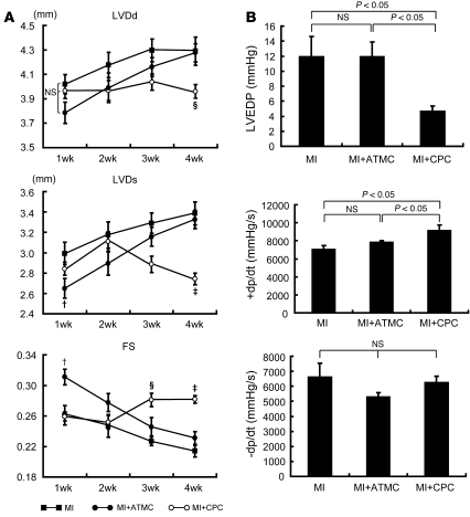
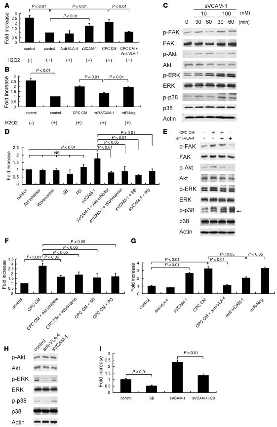
Cardiac progenitor cells are a potential source of cell therapy for heart failure. Although recent studies have shown that transplantation of cardiac stem/progenitor cells improves function of infarcted hearts, the precise mechanisms of the improvement in function remain poorly understood. The present study demonstrates that transplantation of sheets of clonally expanded stem cell antigen 1-positive (Sca-1-positive) cells (CPCs) ameliorates cardiac dysfunction after myocardial infarction in mice. CPC efficiently differentiated into cardiomyocytes and secreted various cytokines, including soluble VCAM-1 (sVCAM-1). Secreted sVCAM-1 induced migration of endothelial cells and CPCs and prevented cardiomyocyte death from oxidative stress through activation of Akt, ERK, and p38 MAPK. Treatment with antibodies specific for very late antigen-4 (VLA-4), a receptor of sVCAM-1, abolished the effects of CPC-derived conditioned medium on cardiomyocytes and CPCs in vitro and inhibited angiogenesis, CPC migration, and survival in vivo, which led to attenuation of improved cardiac function following transplantation of CPC sheets. These results suggest that CPC transplantation improves cardiac function after myocardial infarction through cardiomyocyte differentiation and paracrine mechanisms mediated via the sVCAM-1/VLA-4 signaling pathway.


Figures

Figure 1
Figure 1. Character of CPCs.
(A) CPCs were expanded more than 500 population doublings (P.D.) over a 1-year period. (B and C) Cardiac mRNA and protein expressions in CPCs. (B) Left panels show RT-PCR. Noncontiguous lanes from the same gel were spliced together into a composite band. The thin white line indicates the spliced point. Right panels show Western blot. (C) Immunofluorescent images of CPCs 4 weeks after starting culture under confluent conditions. Scale bars: 100 μm. (D and E) Confocal microscopic images of the infarcted heart 4 weeks after direct injection of RFP+ CPCs. (D) GATA4-expressing RFP+ cells (arrowheads) were recognized in the infarcted region. (E) Some RFP+ cells (arrowheads) expressed sarcomeric α-actinin in the infarcted area. Scale bars: 5 μm.
Figure 2
Figure 2. Effects of CPC sheet transplantation on cardiac function after MI.
(A) Echocardiographic analysis. CPC sheet transplantation inhibited dilatation of LVDd and LVDs and improved FS 3 weeks later. ATMC transplantation inhibited dilatation of LVDs and FS reduction at 1 week, but not afterward. P < 0.05 versus MI or MI plus CPCs (n = 10 per group). P < 0.01 versus MI or MI plus ATMCs (n = 10 per group). §P < 0.05 versus MI or MI plus ATMCs (n = 10 per group). (B) Catheterization analysis at 4 weeks after transplantation. CPC sheet transplantation improved LVEDP and +dp/dt compared with that in the MI or MI plus ATMC groups (n = 5). Data are shown as mean ± SEM.
Figure 3
Figure 3. Immunohistochemical analysis of transplanted hearts.
(A) Masson trichrome staining. The fibrotic area at 4 weeks after transplantation was calculated and is shown in the graph (n = 6). Lower panels show representative images. Scale bars: 1 mm. (B and C) Endothelial cells were identified by immunohistochemical staining with anti-vWF Ab in the border zone of the infarcted hearts 1 week (B) and 4 weeks (C) after transplantation. Lower panels show representative images. The vessel number was quantified and is depicted in the graph (n = 6). HPF, high-power field. Scale bars: 100 μm. Data are shown as mean + SEM.
Figure 4
Figure 4. Cell survival and differentiation of transplanted cells.
(A) Fluorescent microscopic images of infarcted heart 4 weeks after RFP+ CPC sheet transplantation. Left sides of panels show endocardial area. Right sides of panels show epicardial area. Scale bars: 250 μm. (B) Confocal microscopic images of infarcted heart 4 weeks after CPC sheet transplantation (sarcomeric α-actinin, green; RFP, red; Topro, blue; yellow in merged images). Left panel shows normal area. Right panel shows injured area. Arrowheads indicate RFP+ cells. Scale bars: 5 μm. (C) Number of RFP+ cells were quantified and shown in the graph (n = 5). (D and E) Transplanted RFP+ cells expressed sarcomeric α-actinin in a fine striated pattern (D) and formed vessel structures around α-actinin–positive myocardium (E). Nuclei were stained with Topro. Arrowheads indicate vessel structures. Scale bars: 5 μm. (F) Percentages of α-actinin–positive cells or vessel structure–forming cells in existing RFP+ cells were calculated and shown in the graph (n = 5). Data are shown as mean + SEM.
Figure 5
Figure 5. Secreted factor–mediated angiogenesis.
(A) Left panel shows Western blot analysis results using whole-cell lysates of cultured CPCs and ATMCs. Right panel shows the results of sVCAM-1 ELISA using CM from cultured CPCs and ATMCs (n = 3). (B) Western blot analysis results of VCAM-1 and VEGF expression in heart after MI. Normal heart was used as a control. Left panel shows representative images. Right panels show quantification results of VCAM-1 and VEGF expression (n = 3). (C) Scratch-wound assay. CPC-derived CM enhanced endothelial migration (n = 3). Lower panels show representative images (n = 3). Scale bars: 500 μm. (D) CPC-derived CM enhanced endothelial tube formation. Tube length was quantified and is shown in the graph (n = 3). Lower panels show representative images. Scale bars: 500 μm. miR, miRNA. Data are shown as mean + SEM.
Figure 6
Figure 6. sVCAM-1–mediated cardioprotective effects and CPC migration.
(A and B) Cardiomyocyte viability following treatment with H2O2 was measured by MTT assay (n = 3). IgG isotype Abs were used as a control (A). (C) sVCAM-1 induced phosphorylation of FAK, Akt, ERK, and p38 MAPK in a dose-dependent manner. (D) Cardiomyocyte viability following treatment with H2O2 was measured by MTT assay (n = 4). SB, SB2035800; PD, PD98059. (E) CPC-derived CM induced phosphorylation of FAK, Akt, ERK, and p38 MAPK. Anti–VLA-4 Abs inhibited phosphorylation of FAK, Akt, and ERK induced by CPC-derived CM, but not phosphorylation of p38 MAPK. Arrow indicates appropriate size of phosphorylated p38 MAPK. (F) Cardiomyocyte viability following treatment with H2O2 was measured by MTT assay (n = 4). (G and I) CPC migration was measured using the scratch wound assay (n = 3). IgG isotype Ab was used as a control (G). (H) Anti–VLA-4 Abs inhibited phosphorylation of ERK and p38 MAPK of CPCs, but not Akt. Activity of p38 MAPK, but not Akt or ERK, was upregulated by sVCAM-1 treatment. Data are shown as mean + SEM.
Figure 7
Figure 7. The roles of VLA-4 signaling on CPC sheet transplantation-mediated improved cardiac function.
Analysis of cardiac function by echocardiography (A, n = 5) and catheterization (B, n = 5). Anti–VLA-4 Ab treatment inhibited the reduction of LVDd, LVDs, and LVEDP and the improvement of FS and +dp/dt by CPC sheet transplantation. Isotype Ab was used as a control. P < 0.05 versus anti–VLA-4 Abs (n = 5 per group). P < 0.01 versus anti–VLA-4 Abs (n = 5 per group). (C) Masson trichrome staining. The fibrotic area 4 weeks after transplantation was calculated and is shown in the graph (n = 5). Anti–VLA-4 Ab treatment inhibited the reduction of fibrotic area following CPC sheet transplantation. Lower panels show representative images. Scale bars: 1 mm. (D) vWF staining. The number of vWF-positive vessels in the border area was counted and is shown in the graph (n = 5). Anti–VLA-4 Ab treatment inhibited the increased number of vessels in the border area following CPC sheet transplantation. Lower panels show representative images. Scale bars: 100 μm. Nuclei were stained with hematoxylin. (E) RFP staining. The number of RFP-positive cells (brown) was counted and is shown in the graph (n = 5). Anti–VLA-4 Ab treatment decreased the number of RFP+ cells in the infarcted area following CPC sheet transplantation. Lower panels show representative images. Nuclei were stained with hematoxylin. Scale bars: 100 μm. Data are shown as mean + SEM.

Similar articles

Cited by

References

    1. Dimmeler S., Zeiher A.M., Schneider M.D. Unchain my heart: the scientific foundations of cardiac repair. J. Clin. Invest. 2005;115:572–583. - PMC - PubMed
    1. Beltrami A.P., et al. Adult cardiac stem cells are multipotent and support myocardial regeneration. Cell. 2003;114:763–776. doi: 10.1016/S0092-8674(03)00687-1. - DOI - PubMed
    1. Oh H., et al. Cardiac progenitor cells from adult myocardium: homing, differentiation, and fusion after infarction. Proc. Natl. Acad. Sci. U. S. A. 2003;100:12313–12318. doi: 10.1073/pnas.2132126100. - DOI - PMC - PubMed
    1. Matsuura K., et al. Adult cardiac Sca-1-positive cells differentiate into beating cardiomyocytes. J. Biol. Chem. 2004;279:11384–11391. doi: 10.1074/jbc.M310822200. - DOI - PubMed
    1. Messina E., et al. Isolation and expansion of adult cardiac stem cells from human and murine heart. Circ. Res. 2004;95:911–921. doi: 10.1161/01.RES.0000147315.71699.51. - DOI - PubMed

Publication types

MeSH terms

Substances