. 2009 Aug;119(8):2366-78.
doi: 10.1172/JCI38075. Epub 2009 Jul 20.

The muscle-specific microRNA miR-206 blocks human rhabdomyosarcoma growth in xenotransplanted mice by promoting myogenic differentiation

Affiliations

The muscle-specific microRNA miR-206 blocks human rhabdomyosarcoma growth in xenotransplanted mice by promoting myogenic differentiation

Riccardo Taulli et al. J Clin Invest. 2009 Aug.

Abstract

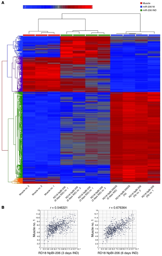
Many microRNAs (miRNAs), posttranscriptional regulators of numerous cellular processes and developmental events, are downregulated in tumors. However, their role in tumorigenesis remains largely unknown. In this work, we examined the role of the muscle-specific miRNAs miR-1 and miR-206 in human rhabdomyosarcoma (RMS), a soft tissue sarcoma thought to arise from skeletal muscle progenitors. We have shown that miR-1 was barely detectable in primary RMS of both the embryonal and alveolar subtypes and that both miR-1 and miR-206 failed to be induced in RMS cell lines upon serum deprivation. Moreover, reexpression of miR-206 in RMS cells promoted myogenic differentiation and blocked tumor growth in xenografted mice by switching the global mRNA expression profile to one that resembled mature muscle. Finally, we showed that the product of the MET proto-oncogene, the Met tyrosine-kinase receptor, which is overexpressed in RMS and has been implicated in RMS pathogenesis, was downregulated in murine satellite cells by miR-206 at the onset of normal myogenesis. Thus, failure of posttranscriptional modulation may underlie Met overexpression in RMS and other types of cancer. We propose that tissue-specific miRNAs such as miR-1 and miR-206, given their ability to modulate hundreds of transcripts and to act as nontoxic differentiating agents, may override the genomic heterogeneity of solid tumors and ultimately hold greater therapeutic potential than single gene-directed drugs.


Figures

Figure 1
Figure 1. miR-1 is poorly expressed in primary tumors, and RMS cells switched to differentiating conditions fail to induce miR-1/miR-206.
(A) Quantitative real-time PCR analysis of mature miR-1 and miR-206 in primary human RMS (A–R) and control muscles (no. 1–4). Mean values (± SD) are from 3 replicates. (B) Increase of expression of mature miR-1/miR-206 in RMS cells and control hMBs in differentiation medium (D, medium with low levels of serum) relative to proliferation medium (P, medium with high levels of serum), measured by real-time PCR. Mean values (± SD) are from 3 independent experiments. (C) Northern blot with miR-206– and miR-1–specific probes on total RNA (5 μg/lane) obtained from the indicated RMS cells grown for 3 days in differentiation medium and from proliferating and differentiating murine satellite cells. Increasing amounts of synthetic miRNAs were used as standards for quantification.
Figure 2
Figure 2. Conditional expression of miR-206 in RMS cells causes reduction of cell proliferation and cell cycle arrest in the G0/G1 phase, increases apoptosis, decreases invasiveness, and enhances myogenic differentiation.
Cells were infected with either the control or the miR-206–expressing vector (NpBI-206AS and NpBI-206, respectively; see Methods) (Tet-off system) and treated (noninduced, NI) or not (induced, IND) with doxycycline (Dox). (A) Proliferation was evaluated for a period of 5 days. The number of cells at day 0 was set at 100%. (B) Cell-cycle distribution of RMS cells in presence/absence of doxycycline was measured by propidium iodide staining and FACS analysis. (C) Apoptosis was measured by Annexin V–allophycocyanin staining and FACS analysis. (D) Invasiveness was evaluated 72 hours after seeding RMS cells in matrigel-coated transwell chambers. (E) MHC expression in RMS cells upon miR-206 induction in medium with high levels of serum compared with controls. Values represent counts of 6 fields for each group normalized against the number of DAPI-positive cells in the same fields (top). Representative immunofluorescence images of induced RD18 cells carrying the NpBI-206AS and the NpBI-206 vector, respectively (bottom). Blue staining was performed with DAPI; red staining was performed with MHC. Original magnification, ×20. (F) Western blot of phospho-pRb, cyclin D1, phospho-p38, myogenin, p21, GFP, and tubulin on noninduced and induced RD18 and RH4 cells (30 μg/lane). All mean values (± SD) are from 3 independent experiments (AE). *P < 0.05; **P < 0.05 (Student’s t test).
Figure 3
Figure 3. Induction of miR-206 shifts the global gene expression profile of RMS cells toward that of muscle.
(A) Unsupervised hierarchical clustering of muscles and NpBI-206 and NpBI-206AS RD18 cells prior to (miR-206 noninduced) and after (miR-206 induced) doxycycline administration (Tet-on system) for the indicated times. Only genes showing a fold change of more than 2 and a t test P value of less than 0.05 were included in the analysis. Red indicates increased expression; blue indicates reduced expression. (B) Pearson correlation of miR-206–expressing RD18 cells (3 and 6 days after induction) compared to normal muscle number 1.
Figure 4
Figure 4. miR-206 arrests growth of RMS xenografts by promoting myogenic differentiation.
(A and B) Continuous expression of pre–miR-206 (green lines) prevents growth of (A) embryonal (RD18) and (B) alveolar (RH4) RMS xenografts. In the Tet-off system, half of the mice (n = 5) were administered drinking water containing 1 mg/ml doxycycline, starting at the time of injection (noninduced), while the rest received water alone (induced). (C and E) Inducible expression of pre–miR-206 arrests growth of (C) RD18 and (E) RH4 xenografts. In the Tet-on system, 5 of 10 mice bearing RMS tumors were given drinking water containing 1 mg/ml of doxycycline, starting on the day indicated by the arrow (green lines, pre–miR-206 induced; black lines, pre–miR-206 noninduced; red lines, AS pre–miR-206 induced [n = 5]). (D and F) miR-206 induction (Tet-on) in advanced RD18 (n = 3) and RH4 (n = 3) tumors is sufficient to block their growth. Doxycycline treatment started on the day indicated by the green arrow. Tumor growth was measured every 3 days, starting when the tumors became palpable (day 0). Bars indicate SEM (AF). (G and H) Immunohistochemical analysis of sections of tumors harvested from doxycycline-treated animals. Ki67-specific antibody was used as a marker for proliferating cells; MHC-specific antibody was used as a marker for differentiated cells. Original magnification, ×20. (I) Representative quantitative real-time PCR analysis of mature miR-206 in RMS xenografts recovered from doxycycline-treated (1 week) and untreated animals.
Figure 5
Figure 5. Met is posttranscriptionally downregulated during myogenic differentiation.
(A) Murine satellite cells grown in proliferation medium (top left panel), differentiate into myotubes when switched to medium with low levels of serum (top right panel). Original magnification, ×20. Representative Northern blot of total RNA (5 μg/lane) from satellite cells (proliferating and at 3 days of differentiation) and adult murine muscles (mouse number 506, 508, and 582), probed for miR-1/miR-206 expression. U6 was used as loading control. Increasing amounts of synthetic miRNAs were used as standards for quantification. (B) Western blot of extracts of satellite cells, either proliferating or at different stages of differentiation (day 1–4), probed for myogenin, MHC, Met, and tubulin as a control. Thirty micrograms of protein extracts were loaded in each lane. (C) Real-time PCR on Met on the same cells. The level of Met transcript in proliferating cells was set at 100%. Mean values (± SD) are from 3 independent experiments.
Figure 6
Figure 6. Met is posttranscriptionally downregulated by miR-206 by direct targeting of its 3′UTR.
(A) Western blot of Met, GFP, and tubulin on protein extracts (30 μg/lane) of murine satellite cells transfected with the Met 3′UTR reporter construct along with a scrambled or miR-206–directed LNA (400 nM) and then switched to differentiation medium for 1 to 2 days. The difference in the kinetics of Met and EGFP downmodulation is most likely due to the long half-life of this form of GFP (stabilized). (B) GFP quantification by FACS analysis on RD18 cells transfected with either a miR-1/miR-206 sensor vector (see Methods) or a point-mutated (MUT) sensor vector along with a scrambled or miR-206–directed LNA (400 nM). The GFP level of control cells was set at 100%. Mean values (± SD) are from 3 independent experiments. (C and D) Western blot on protein extracts of noninduced and induced RD18 and RH4 (C) cells probed for Met, myogenin, and tubulin and (D) RMS xenografts probed for Met and tubulin. Thirty micrograms of protein extracts were loaded in each lane.

Comment in

References

    1. Stefani G., Slack F.J. Small non-coding RNAs in animal development. Nat. Rev. Mol. Cell Biol. 2008;9:219–230. doi: 10.1038/nrm2347. - DOI - PubMed
    1. Bartel D.P. MicroRNAs: genomics, biogenesis, mechanism, and function. Cell. . 2004;116:281–297. doi: 10.1016/S0092-8674(04)00045-5. - DOI - PubMed
    1. Lee R.C., Ambros V. An extensive class of small RNAs in Caenorhabditis elegans. Science. 2001;294:862–864. doi: 10.1126/science.1065329. - DOI - PubMed
    1. Lim L.P., et al. Microarray analysis shows that some microRNAs downregulate large numbers of target mRNAs. Nature. 2005;433:769–773. doi: 10.1038/nature03315. - DOI - PubMed
    1. Baek D., et al. The impact of microRNAs on protein output. Nature. . 2008;455:64–71. doi: 10.1038/nature07242. - DOI - PMC - PubMed

Publication types

MeSH terms