. 2009 Aug;11(8):934-42.
doi: 10.1038/ncb1904. Epub 2009 Jul 20.

Identification of chromosome sequence motifs that mediate meiotic pairing and synapsis in C. elegans

Affiliations

Identification of chromosome sequence motifs that mediate meiotic pairing and synapsis in C. elegans

Carolyn M Phillips et al. Nat Cell Biol. 2009 Aug.

Erratum in

  • Nat Cell Biol. 2009 Sep;11(9):1163

Abstract

Caenorhabditis elegans chromosomes contain specialized regions called pairing centres, which mediate homologous pairing and synapsis during meiosis. Four related proteins, ZIM-1, 2, 3 and HIM-8, associate with these sites and are required for their essential functions. Here we show that short sequence elements enriched in the corresponding chromosome regions selectively recruit these proteins in vivo. In vitro analysis using SELEX indicates that the binding specificity of each protein arises from a combination of two zinc fingers and an adjacent domain. Insertion of a cluster of recruiting motifs into a chromosome lacking its endogenous pairing centre is sufficient to restore homologous pairing, synapsis, crossover recombination and segregation. These findings help to illuminate how chromosome sites mediate essential aspects of meiotic chromosome dynamics.


Figures

Figure 1
Figure 1
The X chromosome PC region. (a) Left two megabases of the X chromosome. Genetic and physical markers used for mapping are indicated. Three deficiencies that remove the PC (meDf2, meDf3, and meDf5) and one that does not (yDf19) were mapped. All PC deficiencies remove pk6142 but not pk6143, indicating breakpoints between 1.46 and 2.07 Mb from the left end. yDf19 removes unc-1 but not pk6141, indicating a breakpoint between 1.06 and 1.17 Mb from the left end. (b-e) HIM-8 immunofluorescence (yellow) in meiotic nuclei from hermaphrodites of the indicated genotypes. Diagrams of the X chromosomes and the mnDp66 duplication, which is required for viability in deficiency homozygotes, are shown. (f-h) HIM-8 immunofluorescence (yellow) was combined with FISH (red) to test for recruitment of HIM-8 to extrachromosomal arrays. Diagram on right indicates the genomic location of the sequences tested in each panel: (f) 539 bp amplicon from cosmid K06A9 on XL. (g) Cluster of TTGGTCAGTGCA repeats from XL. (h) Cluster of 4 HIM-8 recruitment motifs from IIIL recruits HIM-8 (yellow) but not ZIM-1 (green). All images are maximum-intensity projections of deconvolved 3D stacks. Scale bars represent 5 μm.
Figure 2
Figure 2
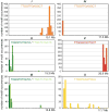
ZIM/HIM-8 recruitment motifs. Distribution of the most abundant ZIM-1 (green), ZIM-2 (red), ZIM-3 (orange), and HIM-8 (yellow) recruitment motif on the six C. elegans chromosomes. Each bin along the X axis represents a 500 kb genomic segment. Note that Y axes show different scales for each chromosome.
Figure 3
Figure 3
Sequence-specific binding by ZIM-2, ZIM-3, and HIM-8 protein fragments. (a) Diagram of ZIM-2, ZIM-3, and HIM-8 showing the full-length proteins (blue), the position of the zinc fingers (ZnF, gold boxes), and the protein fragments expressed in vitro and subjected to SELEX analysis (gray bars). (b) Consensus motifs derived using MEME from the unique sequences identified following four iterations of SELEX (Supplementary Information, Table S3). Protein fragments containing the core ZnF domains of each protein show specific binding to the DNA sequence TTGGC. The randomized region in the library used for SELEX is 21-26 bp, which is significantly larger than the stretch that a 2-finger module can specify. For this reason, oligonucleotides containing two binding sites are more likely to be co-precipitated with tagged proteins than sequences containing a single site (E. Rebar, pers. comm.). A C-terminal fragment of HIM-8, including the ZnF domains, specifically binds to the same sequence identified by recruitment of HIM-8 to extrachromosomal arrays.
Figure 4
Figure 4
ZIM/HIM-8 recruiting arrays associate with nuclear envelope components. (a-c) Combined FISH detection of extrachromosomal arrays (red) and immunolocalization of HIM-8 or ZIM-3 (yellow) and ZYG-12:GFP (green). HIM-8 (a) and ZIM-3 (b) recruiting arrays interact with a large patch of ZYG-12 at the nuclear envelope. The array shown in (c) does not recruit HIM-8 or any of the ZIM proteins, and does not colocalize with ZYG-12. Arrows indicate clear examples of co-localization (or lack thereof, in c). All images are maximum-intensity projections of deconvolved 3D stacks. Scale bars represent 5 μm.
Figure 5
Figure 5
HIM-8 recruitment motifs are sufficient for PC function. (a) Integration of a HIM-8 recruiting array (see Fig. 1f) onto meDf2, a PC-deficient X chromosome. (b) Hermaphrodite homozygous for meDf2 has unsynapsed X chromosomes in most pachytene nuclei, visualized as axial elements marked by HTP-3 (red) lacking the central element protein SYP-1 (green). Arrows indicate examples of unsynapsed chromosomes. (c) Most meiotic nuclei in meDf2 ieIs5 hermaphrodites are fully synapsed. (d, e) Stabilization of pairing in the absence of synapsis (syp-2 RNAi) is examined by performing FISH to the integrated array (red). In meDf2 ieIs5 oocytes (d) the arrays are paired, as indicated by only a single region of FISH staining. In him-8; meDf2 ieIs5 oocytes (e) the arrays are unpaired, indicating that pairing between integrated arrays, like that seen between endogenous PCs, is him-8 dependent. (f, g) Oocytes at diakinesis in meDf2 and meDf2 ieIs5 hermaphrodites. FISH probes to the center (yellow) and right end (red) identify the X chromosomes. Arrows indicate non-recombinant (univalent) and recombinant (bivalent) X chromosomes in meDf2 and meDf2 ieIs5 hermaphrodites, respectively. (h) Quantification of recombinant X chromosomes. (i) Quantification of males produced by self-fertilizing hermaphrodites of the indicated genotypes. Scale bars represent 5 μm.
Figure 6
Figure 6
ZIM/HIM-8 proteins can interchangeably support PC function. (a-d) Oocytes at diakinesis from [zim-2], [zim-2; ieIs12], [him-8], and [him-8; ieIs14] hermaphrodites. FISH probes to the 5S rDNA (red in a, b) and an X-chromosome repeat (red in c, d) were used to identify chromosomes V and X, respectively. In animals carrying chromosomal insertions of ZIM-2/HIM-8 binding sites ieIs12 (b) and ieIs14 (d), the chromosome-specific probes localize to a single bivalent, which is also marked by a FISH probe to λ DNA (green), indicating that the insertion of binding sites restored crossover recombination on chromosome V in zim-2 animals and the X chromosome in him-8 animals. (e) Quantification of bivalent X (red) and V (orange) chromosomes. (f) Quantification of males produced by self-fertilizing hermaphrodites of the indicated genotypes. (g,h) Arrays of binding sites (red) inserted into two different chromosomes (ieIs12/+; ieIs14/+ and ieIs13/+; ieIs15/+) were assayed for their ability to promote stable pairing between non-homologous chromosomes in the absence of synapsis (syp-2 RNAi). (i) Synapsis was analyzed in animals heterozygous for two different insertions (ieIs13/+; ieIs15/+) by immunostaining of SC components. Nuclei containing unsynapsed chromosomes, visualized as segments positive for the axial element protein HTP-3, (red) but lacking transverse filament proteins including SYP-1 (green), usually contain integrated arrays (blue) that are paired and synapsed with each other, indicating non-homologous synapsis between chromosomes V and X (arrows). In contrast, nuclei with fully synapsed chromosomes often contained unpaired arrays (blue), indicating that all chromosomes are likely synapsed with their appropriate homologs (arrowheads). Scale bars represent 5 μm.

Comment in

Similar articles

Cited by

References

    1. McKim KS, Howell AM, Rose AM. The effects of translocations on recombination frequency in Caenorhabditis elegans. Genetics. 1988;120:987–1001. - PMC - PubMed
    1. McKim KS, Peters K, Rose AM. Two types of sites required for meiotic chromosome pairing in Caenorhabditis elegans. Genetics. 1993;134:749–768. - PMC - PubMed
    1. Villeneuve AM. A cis-acting locus that promotes crossing over between X chromosomes in Caenorhabditis elegans. Genetics. 1994;136:887–902. - PMC - PubMed
    1. Zetka M, Rose A. The genetics of meiosis in Caenorhabditis elegans. Trends Genet. 1995;11:27–31. - PubMed
    1. Zetka MC, Rose AM. The meiotic behavior of an inversion in Caenorhabditis elegans. Genetics. 1992;131:321–332. - PMC - PubMed

Publication types

MeSH terms

Substances