. 2009 Aug;12(8):1020-7.
doi: 10.1038/nn.2371. Epub 2009 Jul 20.

Balanced gene regulation by an embryonic brain ncRNA is critical for adult hippocampal GABA circuitry

Affiliations

Balanced gene regulation by an embryonic brain ncRNA is critical for adult hippocampal GABA circuitry

Allison M Bond et al. Nat Neurosci. 2009 Aug.

Abstract

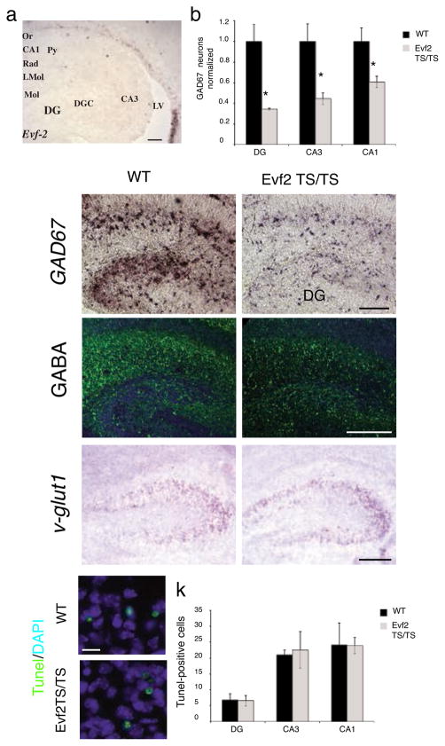
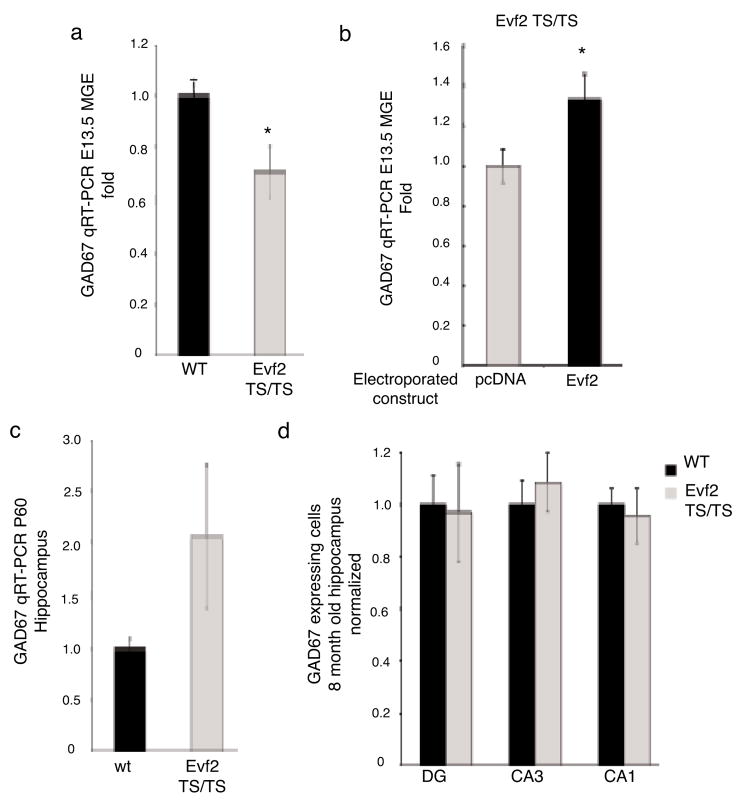
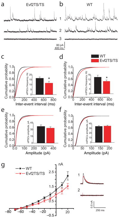
Genomic studies demonstrate that, although the majority of the mammalian genome is transcribed, only about 2% of these transcripts are code for proteins. We investigated how the long, polyadenylated Evf2 noncoding RNA regulates transcription of the homeodomain transcription factors DLX5 and DLX6 in the developing mouse forebrain. We found that, in developing ventral forebrain, Evf2 recruited DLX and MECP2 transcription factors to important DNA regulatory elements in the Dlx5/6 intergenic region and controlled Dlx5, Dlx6 and Gad1 expression through trans and cis-acting mechanisms. Evf2 mouse mutants had reduced numbers of GABAergic interneurons in early postnatal hippocampus and dentate gyrus. Although the numbers of GABAergic interneurons and Gad1 RNA levels returned to normal in Evf2 mutant adult hippocampus, reduced synaptic inhibition occurred. These results suggest that noncoding RNA-dependent balanced gene regulation in embryonic brain is critical for proper formation of GABA-dependent neuronal circuitry in adult brain.


Figures

Figure 1
Figure 1. Evf2TS/TS mice have increased Dlx5 and Dlx6 expression in the embryonic brain
a. Schematic of targeting a triple polyadenylation transcription stop site into Evf exon1, truncating Evf2 transcripts from 3.7 kb to 101 bp (Evf2TS), but not Evf1, which is transcribed starting from exon3. Truncated Evf2TS transcript (101 bp) completely lacks the ultraconserved ei region. Only the distance from TS insertion to the Dlx5 (~10.2 kb) and Evf1 (~10.4 kb) transcription start sites are shown to scale. b. ES cells used for making Evf2TS mice contain correctly targeted transcription stop into Evf exon1, as verified by Southern analysis. c–h. RNA in situ hybridization of E13.5 coronal sections of wildtype (wt) and Evf2TS/TS mutant telencephalon, probed with anti-sense Evf2 (c, d), anti-sense Evf1 (e, f), or anti-sense Dlx5 (g, h). i. Quantitative real-time RT-PCR analysis of E13.5 MGE from wt and Evf2TS/TS mutants. Error bars: S.E.M. Statistical analysis: Mann Whitney U-test, p<.05: Evf2=0.0055, Dlx5=0.044, Dlx6=0.0055; n= 4 for wt, n=6 for Evf2TS/TS. j. Quantitative real-time RT-PCR analysis of E12.5 Evf2TS/TS mutant MGE electroporated with: control (pcDNA, 2μg), 1 μg pcDNA-Evf2 (+1 μg pcDNA), and 2μg pcDNA-Evf2. Error bars: S.E.M. Statistical analysis, ANOVA Dunnett’s two-sided test, p<.05 values: Dlx5, (0μg,1μg Evf2=0.05, 0μg,2μg Evf2=0.001), ANOVA Tukey test, p<.05 values, Dlx6 (0μg,2μg Evf2=0.041, 1μg Evf2, 2μg Evf2= .033); n=4. * p < 0.05; t-test, Mann Whitney, or ANOVA
Figure 2
Figure 2. Loss of Evf2 affects DLX and MECP2 binding to Dlx 5/6 intergenic enhancers in embryonic day 13.5 medial ganglionic eminence
a. Schematic showing the region in mouse chromosome 6qA1 surrounding Dlx5/6 and Evf1/2. Quantitative chromatin immunoprecipitation of E13.5 MGE from wt (black bars) and Evf2TS/TS mutants (gray bars) using primers 1–6 across the Dlx 5/6 region, and antibodies to: b. anti-pan DLX, p <.05 values: 3=0.025, 5=0.025, c. anti-MECP2, p<.05 values: 2=0.025, 3=0.025, 5=0.025, d. anti-HDAC1, p <.05 values: 5=0.025, 6=0.025. qChIP-PCR was performed on optimized primer sets using previously defined primer sets (1=15, 2=24, 4=27) and newly defined sets for ei (3- red), eii (5-red) and external primer (6). Primer 2 (green) was identified as a MECP2/HDAC binding site for transcriptional repression in adult cortex. Error bars: S.E.M. Statistical analysis: Mann Whitney U-test. * p < 0.05 Mann Whitney U-test. The schematic in (a) is reversed from that shown for Evf2 transcription-stop insertion (Fig 1) in order to align with previously published primers.
Figure 3
Figure 3. Evf2 does not affect DLX or MECP2 nuclear localization
a. Western Blot analysis of DLX2 protein after transfection of a construct containing Dlx2 cDNA and/or Evf2 cDNA. Transfection was performed into C17 mouse neural stem cells in the presence or absence of Evf2 RNA, showing equal levels of DLX2 protein. That Evf2 RNA is stable after transfection was verified by RT-PCR (not shown). b. Western analysis of Evf2TS/TS and wt E13.5 ganglionic eminence extracts probed with anti-DLX2 specific antibody (gift of David Eisentstat) shows that loss of Evf2 does not affect DLX2 protein levels. c–h. Pan-anti-DLX antibody (green) stains nuclei counterstained with DAPI (blue) in both wt and Evf2TS/TS E13.5 MGE. MGE, medial ganglionic eminence; VZ, ventricular zone; SVZ, subventricular zone. c, f: scale bar= 200μm; d, g: scale bar=20μm; e, h: scale bar= 10μm. i–j. Immunohistochemistry using anti-MECP2 antibodies (green) and DAPI (blue nuclei) shows similar nuclear localization in both wt and Evf2TS/TS MGE cells, scale bar=20μm.
Figure 4
Figure 4. GABAergic interneuron loss in the P2 hippocampus and dentate gyrus of Evf2TS/TS mutant mice
a. RNA in situ hybridization analysis of Evf2 expression in the subventricular zone and cells lining the lateral ventricle as they migrate to the hippocampus and dentate gyrus. scale bar=130μm; Or, oriens; Py, pyramidal; Rad, radiatum; LMol, lacunosum molecular; Mol, molecular layer of the dentate gyrus; DGC, dentate granule cell layer; DG, dentate gyrus; LV, lateral ventricle. b. Quantification of the number of GAD67-expressing GABAergic interneurons in the dentate gyrus (DG), and hippocampal CA1 and CA3 regions. Error bars: S.E.M. Statistical analysis: Mann Whitney U-test, p =.025 for DG, CA3, CA1; n= 3 for wt, n=3 for Evf2TS/TS. c–d. RNA in situ hybridization of GAD67 expressing interneurons in the hippocampus, scale bar=135μm e–f. Immunohistochemistry, anti-GABA antibodies (green) and DAPI (blue nuclei), scale bar= 200 μm. g–h. RNA in situ hybridization of v-glut1 expressing neurons in the hippocampal CA3 region shows similar expression in wt and Evf2TS/TS, scale bar=80μm. i–j. Immunohistochemistry using the apoptosis TUNEL assay (green) and DAPI (blue nuclei) in the hippocampal CA3 region, scale bar= 10μm. k. Quantification of the number of TUNEL-positive cells in the dentate gyrus (DG), and hippocampal CA1 and CA3 regions shows no difference in cell death between wt and Evf2TS/TS mutants. Error bars: S.E.M. Statistical analysis: Student’s t-test, p >0.05 for DG, CA3, CA1; n= 3 for wt, n=3 for Evf2TS/TS. * p < 0.05; t-test or Mann Whitney U-test
Figure 5
Figure 5. Evf2 trans-positively regulates GAD67 expression in E13.5 MGE, but not adult hippocampus
a. Quantitative real-time RT-PCR analysis of GAD67 in E13.5 MGE from wt and Evf2TS/TS mutants. Error bars: S.E.M. Statistical analysis: Student’s t-test, p=0.031; n= 3 for wt, n=5 for Evf2TS/TS. b. Quantitative real-time RT-PCR analysis of GAD67 in E12.5 Evf2TS/TS mutant MGE electroporated with: control (pcDNA, 2μg), and 2μg pcDNA-Evf2. Error bars: S.E.M. Statistical analysis: Student’s t-test, p=0.0375; n=4. c. Quantitative real-time RT-PCR analysis of GAD67 in P60 hippocampus from wt and Evf2TS/TS mutants. Error bars: S.E.M. Statistical analysis: Student’s t-test, p=0.031; n=2 for wt, n=2 for Evf2TS/TS. d. Quantification of the number of GAD67-expressing GABAergic interneurons in 8-month old dentate gyrus (DG), and hippocampal CA3 and CA1 regions. Error bars: S.E.M. Statistical analysis: Student’s t-test, p>0.05 for DG, CA3, CA1; n=3 for wt, n=3 for Evf2TS/TS. * p < 0.05; t-test
Figure 6
Figure 6. GABAergic synaptic inhibition is reduced in CA1 layer of the adult hippocampus of Evf2TS/TS mutant mice
(a–b) Representative traces of sIPSCs (1) and mIPSCs (2) from CA1 pyramidal cells of wildtype (WT) and Evf2TS/TS mice. Picrotoxin (PTX) 0.25 mM blocks all IPSC activity in Evf2TS/TS and WT mice (3). (c–d) Averaged cumulative probability plots of sIPSC and mIPSC inter-event intervals from CA1 pyramidal cells (Evf2TS/TS, red; WT, black; Kolmogorov-Smirnov test, p<0.001). sIPSCs and mIPSCs event frequency (insert) in adult and old Evf2TS/TS mice (sIPSC adult: 11.75±1.73 Hz, sIPSC old: 9.17±0.71; mIPSCs adult: 10.65±1.05 Hz, mIPSCs old: 6.5±0.83 Hz) than in control WT mice (sIPSC adult: 16.66±1.26 Hz, sIPSC old: 12.62±0.92 Hz; mIPSCs adult: 14.16±1.67 Hz, mIPSCs old: 9.06±0.77 Hz). (e–f) Averaged cumulative probability plots of sIPSCs and mIPSCs amplitude. Evf2TS/TS, red; WT, black, sIPSC adult: 33.6±2.7 pA, sIPSC old: 48.50±3.06; mIPSCs adult: 35.26±1.41 pA, mIPSCs old: 34.87±3.76) and WT mice (sIPSC adult: 37.4±3.2 pA, sIPSC old: 53.78±3.98; mIPSCs adult: 34.54±1.64 pA, mIPSCs old: 37.03±4.63). (g) The I–V plot of evoked IPSCs of GABAergic inhibition in Evf2TS/TS mice (p<0.01; paired t-test). IPSC current was normalized to the amplitude of evoked EPSC in ACSF containing no drugs to suppress excitatory synaptic activity. (1) Representative IPSCs recordings at +20 mV in Evf2TS/TS (red) and WT (black) mice (average n=5) (2) PTX 0.25 mM blocks evoked IPSCs in CA1 neurons from both WT and Evf2TS/TS mice. Error bars: S.E.M.; * p < 0.05; t-test.

Similar articles

Cited by

References

    1. Prasanth KV, Spector DL. Eukaryotic regulatory RNAs: an answer to the ‘genome complexity’ conundrum. Genes Dev. 2007;21:11–42. - PubMed
    1. Mattick JS. A new paradigm for developmental biology. J Exp Biol. 2007;210:1526–1547. - PubMed
    1. Shamovsky I, Nudler E. Gene control by large noncoding RNAs. Sci STKE. 2006;2006:pe40. - PubMed
    1. Feng J, et al. The Evf-2 noncoding RNA is transcribed from the Dlx-5/6 ultraconserved region and functions as a Dlx-2 transcriptional coactivator. Genes Dev. 2006;20:1470–1484. - PMC - PubMed
    1. Zerucha T, et al. A highly conserved enhancer in the Dlx5/Dlx6 intergenic region is the site of cross-regulatory interactions between Dlx genes in the embryonic forebrain. J Neurosci. 2000;20:709–721. - PMC - PubMed

Publication types

MeSH terms

LinkOut - more resources