. 2009 Aug;5(8):551-8.
doi: 10.1038/nchembio.202.

Enzymatic transition states and dynamic motion in barrier crossing

Affiliations

Enzymatic transition states and dynamic motion in barrier crossing

Steven D Schwartz et al. Nat Chem Biol. 2009 Aug.

Abstract

What are the atomic motions at enzymatic catalytic sites on the timescale of chemical change? Combined experimental and computational chemistry approaches take advantage of transition-state analogs to reveal dynamic motions linked to transition-state formation. QM/MM transition path sampling from reactive complexes provides both temporal and dynamic information for barrier crossing. Fast (femtosecond to picosecond) dynamic motions provide essential links to enzymatic barrier crossing by local or promoting-mode dynamic searches through bond-vibrational space. Transition-state lifetimes are within the femtosecond timescales of bond vibrations and show no manifestations of stabilized, equilibrated complexes. The slow binding and protein conformational changes (microsecond to millisecond) also required for catalysis are temporally decoupled from the fast dynamic motions forming the transition state. According to this view of enzymatic catalysis, transition states are formed by fast, coincident dynamic excursions of catalytic site elements, while the binding of transition-state analogs is the conversion of the dynamic excursions to equilibrated states.


Figures

Figure 1
Figure 1
Transition-state stabilization compared to free energy during an enzymatic reaction. (a) The transition-state stabilization proposal suggests a thermodynamic equilibrium in which tight binding of reactants (S) to the enzyme (E) at the transition state (‡) is proportional to enzymatic rate enhancement (kenz/kchem). (b) Distinct timescales define protein organization leading to transition-state formation, barrier crossing and product release. Reaction barrier crossing is typically shown as taking 50–100 fs, whereas the yellow bar shows a transition-state lifetime of ~5 fs. There is no evidence for a high-energy intermediate, as indicated by the shallow barrier with the question mark.
Figure 2
Figure 2
Transition-state structures of PNP and the effect of remote mutations. (a) The molecular electrostatic potentials are shown at the van der Waals surface as obtained from KIE and computational analysis of human PNP. The stick figures shown above correspond to the potential surfaces. The Michaelis constant for the substrate inosine is contrasted to the dissociation constant for the transition-state mimic DADMe-ImmH. Adapted with permission from ref. . Copyright 2004 American Chemical Society. (b) Comparison of the reaction coordinates for inosine arsenolysis catalyzed by human (HsPNP), bovine (BtPNP) and K22E:H104R chimeric human (Bt-HsPNP) PNPs. The highest energetic points are the chemical transition states. The first barrier represents dissociation of the N-ribosidic bond, and the second barrier corresponds to the attack of the nucleophile. A ribocation species is between these transition states (dashed blue line). BtPNP has an early dissociative transition state (black). The HsPNP transition state features full ribocation character. The transition state for Bt-HsPNP involves some bond formation to the oxygen nucleophile. Reprinted with permission from ref. . Copyright 2008 American Chemical Society. (c) The human PNP trimer, with Lys22 and His104 residues highlighted and the catalytic sites filled with Immucillin-H, a transition-state analog. The Phe159 loop interacts with the catalytic site on the neighboring subunit. The catalytic sites are located near the subunit interface. Lys22 and His104 are located at 45 Å and 25 Å away from the enzyme active site, respectively. The structure is taken from Protein Data Bank accession number 1PF7. (d) The active site residues near the bound transition-state analog Immucillin-G and the phosphate nucleophile. The ImmG:O59, ImmG:N49, H257:Nd and HPO4:Op atoms are highlighted in red. (e) Active site residues in contact with Immucillin-G and phosphate. The dashed red bonds represent oxygens in close proximity in the transition state and constitute the three-oxygen stack.
Figure 3
Figure 3
BIEs for [5′-3H]inosine, Immucillin-H and DADMe-Immucillin-H to human PNP. The KIE for inosine was measured for the arsenolysis reaction, and the BIE was measured with sulfate as an unreactive phosphate analog. Reprinted with permission from ref. . Copyright 2008 American Chemical Society.
Figure 4
Figure 4
Transition path sampling studies of catalysis by lactate dehydrogenase. (a) Schematic drawing of the shooting algorithm. Starting with a single reactive trajectory, an ensemble of reactive paths are generated by randomly choosing a time slice on which a perturbation of each momentum, chosen from a Boltzmann distribution, is applied. (b) A correlation function analysis of the resultant trajectories demonstrates that trajectories decorrelate after approximately 30 shooting moves. Panels a, b reprinted with permission from ref. . Copyright 2005 American Chemical Society. (c) A representative commitment probability for a reactive trajectory for the reaction catalyzed by lactate dehydrogenase. If one views the time in the transition-state region as the time spent with a probability of barrier crossing near 0.5, only a fraction of a femtosecond is spent in the transition-state region. (d) Overlays of atomic coordinates from a variety of paths through phase space to the transition state reveal that although many nonidentical geometries are sampled, the transition-state features are almost superimposable. (e,f) The monomeric unit of LDH and the promoting vibration that is found to be an integral part of the reaction coordinate. The promoting vibration is formed from amino acids extending from one edge of the protein backbone through the active site and to the opposite edge of the enzyme (f). Panels c–f reprinted from ref. . Copyright (2007) National Academy of Sciences U.S.A.
Figure 5
Figure 5
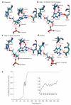
Steps in the phosphorolysis of guanosine by PNP. (a) PNP compresses a three-oxygen stack to polarize the ribosidic bond and create a ribocation. (b) Creation of the ribocation, with full separation of the guanine leaving group. (c) Migration of the ribose ring toward the phosphate nucleophile. (d) Formation of the product with a new bond to form α-d-ribose 1-phosphate. (e) The complex nature of this transition-state barrier process is shown through the committor distribution. The process involves a substantially longer lifetime than the reaction catalyzed by lactate dehydrogenase. The transition-state lifetime (0.5 committor probability) is approximately 10 fs. This time is too short for equilibration through the protein. The snapshots shown here can be viewed as a movie (see also Fig. 6). Reprinted from ref. . Copyright (2008) National Academy of Sciences USA.
Figure 6
Figure 6
The starting point snapshot from a movie demonstrating protein and reactant motion for barrier crossing in human PNP by transition-path sampling. The full movie is available as Supplementary Movie 1. The starting point shows guanosine and phosphate as reactants, with the distances (in Å) to protein groups involved in barrier crossing. Reactants are treated by QM and the protein by MM. The ribosyl group is centered in the figure with the 4′ ring oxygen in red. The cluster of blue atoms at 2 o’clock is the edge-on view of the guanine leaving group. Multiple interactions in phosphate activation are not shown here, but all protein and reactant atoms were included in the QM/MM calculations. Reprinted from ref. . Copyright (2008) National Academy of Sciences USA.

References

    1. Pauling LC. Molecular architecture and biological reactions. Chem. Eng. News. 1946;24:1375–1377.
    1. Wolfenden R. Transition state analogues for enzyme catalysis. Nature. 1969;223:704–705. - PubMed
    1. Hammes-Schiffer S, Benkovic SJ. Relating protein motion to catalysis. Annu. Rev. Biochem. 2006;75:519–541. - PubMed
    1. Gao J, et al. Mechanisms and free energies of enzymatic reactions. Chem. Rev. 2006;106:3188–3209. - PMC - PubMed
    1. Schramm VL. Enzymatic transition state theory and transition state analogue design. J. Biol. Chem. 2007;282:28297–28300. - PubMed