. 2009 Jul 21;4(7):e6298.
doi: 10.1371/journal.pone.0006298.

Hyperactivation of DNA-PK by double-strand break mimicking molecules disorganizes DNA damage response

Affiliations

Hyperactivation of DNA-PK by double-strand break mimicking molecules disorganizes DNA damage response

Maria Quanz et al. PLoS One. .

Abstract

Cellular response to DNA damage involves the coordinated activation of cell cycle checkpoints and DNA repair. The early steps of DNA damage recognition and signaling in mammalian cells are not yet fully understood. To investigate the regulation of the DNA damage response (DDR), we designed short and stabilized double stranded DNA molecules (Dbait) mimicking double-strand breaks. We compared the response induced by these molecules to the response induced by ionizing radiation. We show that stable 32-bp long Dbait, induce pan-nuclear phosphorylation of DDR components such as H2AX, Rpa32, Chk1, Chk2, Nbs1 and p53 in various cell lines. However, individual cell analyses reveal that differences exist in the cellular responses to Dbait compared to irradiation. Responses to Dbait: (i) are dependent only on DNA-PK kinase activity and not on ATM, (ii) result in a phosphorylation signal lasting several days and (iii) are distributed in the treated population in an "all-or-none" pattern, in a Dbait-concentration threshold dependant manner. Moreover, despite extensive phosphorylation of the DNA-PK downstream targets, Dbait treated cells continue to proliferate without showing cell cycle delay or apoptosis. Dbait treatment prior to irradiation impaired foci formation of Nbs1, 53BP1 and Rad51 at DNA damage sites and inhibited non-homologous end joining as well as homologous recombination. Together, our results suggest that the hyperactivation of DNA-PK is insufficient for complete execution of the DDR but induces a "false" DNA damage signaling that disorganizes the DNA repair system.


Conflict of interest statement

Competing Interests: C. Agrario: employee of DNA Therapeutics. M. Quanz: recipient of a PhD fellowship cofinanced by DNA Therapeutics. M. Dutreix and J-S. Sun: cofounders of DNA Therapeutics.

Figures

Figure 1
Figure 1. DNA-PK activation and H2AX phosphorylation by Dbait treatment.
(A) Various 32P-labeled Dbait molecules (32H, 24H, 16H, 8H), were incubated with increasing amounts (0-320 ng/ml) of Hep-2 nuclear extract and analyzed on polyacrylamide gels. Asterisks indicate one or two dimer shift. Antibodies against Ku80 were added prior to migration to confirm by supershift that the bands indicated by asterisks contained Ku80 protein: Lane 1, 32H; lane 2, 32H + anti-Ku80; lane 3, 32H + nuclear extract; lane 4, 32H + nuclear extract + anti-Ku80. The diamond indicates the supershifted band of Dbait–Ku complexes bound by anti-Ku80. (B) Stimulation of DNA-PK kinase activity by 20 mM of various Dbait molecules was measured in 1.5 µg HEp-2 nuclear extract. When indicated, inhibitors NU7026 (NU) and wortmannin (Wm) were preincubated with extract 5 min prior to 32H addition (hatched). Data represent the mean value and standard deviation of at least three independent experiments. (C-E) Cells were transfected (5 h) with different Dbait molecules as indicated and irradiated (10 Gy) or not. If not indicated otherwise, proteins were extracted 1 h after end of treatment for western blot analysis. (C) HEp-2 cell lysates were probed for γ-H2AX and β-actin. (D) Immunoblot of phosphorylated DNA-PKcs; total DNA-PKcs was revealed on a separate membrane. (E) V3 (DNA-PKcs-/-), KA4 (DNA-PKcs kinase deficient) and F18 cells (DNA-PKcs proficient) were treated as indicated and the lysates were probed for γ-H2AX and total H2AX.
Figure 2
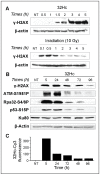
Figure 2. Kinetics of the 32Hc induced response.
(A) MRC-5 cells were transfected or irradiated at time 0 and then extracted for western blot analysis of γ-H2AX and β-actin at the indicated times. (B-C) To monitor the kinetics of DNA-PK target protein phosphorylation and 32Hc elimination, cells were transfected with Cy3-conjugated 32Hc and (B) one part of each sample was processed for SDS-PAGE and hybridized with antibodies against β-actin, Ku80, and phosphorylated p53, Rpa32 and H2AX. Phosphorylated ATM was revealed on a separate gel due to its higher molecular weight. (C) In parallel, the other part of the sample was analyzed by flow cytometry for the mean Cy3-fluorescence.
Figure 3
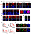
Figure 3. Phosphorylation pattern of DNA damage signaling proteins in 32Hc treated cells.
(A) Immunostaining of γ-H2AX in MRC-5 cells after 10 Gy IR (left panel) and in various cell lines after 32Hc treatment. Cells were fixed 2 h after IR and 1 h after the end of transfection. (B-D) Phosphorylation pattern of different DDR proteins in MRC-5 cells after 32Hc treatment. Cells were immunostained with antibodies against γ-H2AX, Chk2-T68P, Chk1-S345P, Rpa32-S4/8P, ATM-S1981P, Nbs1-S343P, p53-S15P as indicated. (B) Interphase cells. (C) Co-localization of γ-H2AX and DNA (D) M-Phase cells. (E) 32Hc treated M059K and M059J cells were probed for ATM-S1981P. (F) Flow cytometry analysis of not-treated (NT, gray), irradiated (5 Gy, upper left panel) or 32Hc transfected MRC-5 cells with immunostaining for the indicated phosphoproteins. Percentages reflect the proportion of the “positive” cells after 32Hc treatment. (G) Co-staining of γ-H2AX and Rpa32-S4/8P (upper panel) or Chk1-S345P (lower panel). Arrows indicate γ-H2AX positive cells that are negative for the other phosphoprotein. Scale bar: 20 µm.
Figure 4
Figure 4. Characteristics of γ-H2AX positive cells.
Flow cytometry analysis of 32Hc treated MRC-5 cells co-stained for various phosphorylated proteins and DNA content (A) Distribution of the population stained with γ-H2AX. (B) quantification of the cell cycle distribution of the total cell population and the γ-H2AX, P-p53 or P-Chk2 “positive” subpopulation of the indicated proteins. (C) Percentage of γ-H2AX positive cells in the population (as quantified by flow cytometry) after transfection with varying amounts of 32Hc. (D) Dotplot of cells transfected with Cy5-conjugated 32Hc and immunostained for γ-H2AX (upper panel). Lower panel: 32Hc-Cy5 distribution in the cell population (black) with superposed subpopulations gated for γ-H2AX negative (gray) and positive (red) cells.
Figure 5
Figure 5. Effect of 32Hc on cell cycle and viability.
(A) Proliferation rates of untreated (squares), 8H (circles) or 32Hc (triangles) treated MRC-5 (left panel) and HeLa (right panel) cells. Error bars represent SD. (B) Flow cytometric cell cycle analysis of untreated, 8H or 32Hc transfected and irradiated (10 Gy) MRC-5 cells. DNA was stained with propidium iodide. Cells were rediluted once after 24 h. (C) DNA synthesis was analyzed in HEp-2 cells by pulsing cells with BrdU at 24 h after 32Hc treatment: Left Panel: cells stained with γ-H2AX antibodies; Right Panel: cells stained with BrdU antibodies (arrows indicate γ-H2AX positive cells). (D) The cell cycle rate was estimated by microscopy analysis of HEp-2 cells' nuclei pulsed with BrdU 1h after transfection and grown for 24 h before fixation and γ-H2AX, BrdU co-immunostaining (see material and methods). In situ measurement of DNA content and the pattern of BrdU labeling indicated cells having completed a cell cycle.
Figure 6
Figure 6. Effect of 32Hc on IRIF formation and DNA repair.
(A) Inhibition of radiation-induced plasmid integration. Cells were transfected with 2 µg circular plasmid and 2 µg of 32Hc or 8H. The percentage of purR cells having integrated the plasmid was calculated after 5 Gy irradiation (black columns) or without irradiation (grey columns) by dividing the number of colony-forming units in the presence of puromycin by the number of colony-forming units in the absence of puromycin for each transfection condition. The values indicated are means of at least three independent experiments. NU, NU7026; Wm, wortmannin. (B) Immunodetection of Rad51 (green) and γ-H2AX (red) in irradiated MRC-5 cells (10 Gy). Cells were fixed 3 h after the end of transfection and IR. DNA was stained with DAPI. Scale bar: 20 µm. Histograms show the quantification (200 cells counted) of the number of foci in γ-H2AX-positive (black) or negative (white) cells. The median values were four foci per γ-H2AX-positive cells and 11 foci per γ-H2AX-negative cells (C) Inhibition of HR. Human RG37 cells containing a tandem repeat of two inactive cassettes coding for EGFP (one of which contains a cleavage site for the meganuclease I-SceI) were transfected with HA-I-SceI expression vector and co-transfected with 8H, 32Hc or not (NT). Recombinant cells express functional EGFP and were measured by flow cytometry. The efficiency of transfection of HA-tagged-I-SceI was followed using an anti-HA antibody and was comparable for all three conditions.

References

    1. Lett JT. Damage to DNA and chromatin structure from ionizing radiations, and the radiation sensitivities of mammalian cells. Prog Nucleic Acid Res Mol Biol. 1990;39:305–352. - PubMed
    1. Ward JF. The yield of DNA double-strand breaks produced intracellularly by ionizing radiation: a review. Int J Radiat Biol. 1990;57:1141–1150. - PubMed
    1. Rouse J, Jackson SP. Interfaces between the detection, signaling, and repair of DNA damage. Science. 2002;297:547–551. - PubMed
    1. Zhou BB, Elledge SJ. The DNA damage response: putting checkpoints in perspective. Nature. 2000;408:433–439. - PubMed
    1. Aten JA, Stap J, Krawczyk PM, van Oven CH, Hoebe RA, et al. Dynamics of DNA double-strand breaks revealed by clustering of damaged chromosome domains. Science. 2004;303:92–95. - PubMed

Publication types