. 2009 Aug;238(8):1936-50.
doi: 10.1002/dvdy.22025.

High resolution map of Caenorhabditis elegans gap junction proteins

Affiliations

High resolution map of Caenorhabditis elegans gap junction proteins

Zeynep F Altun et al. Dev Dyn. 2009 Aug.

Erratum in

Abstract

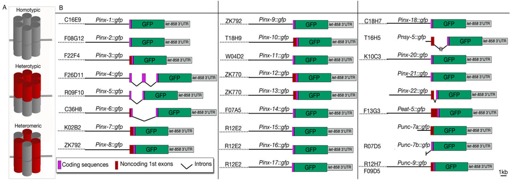
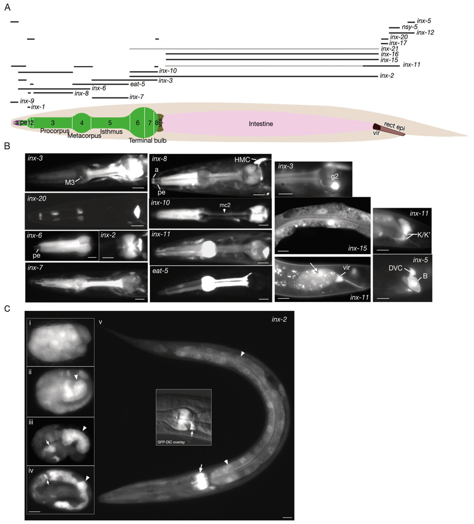
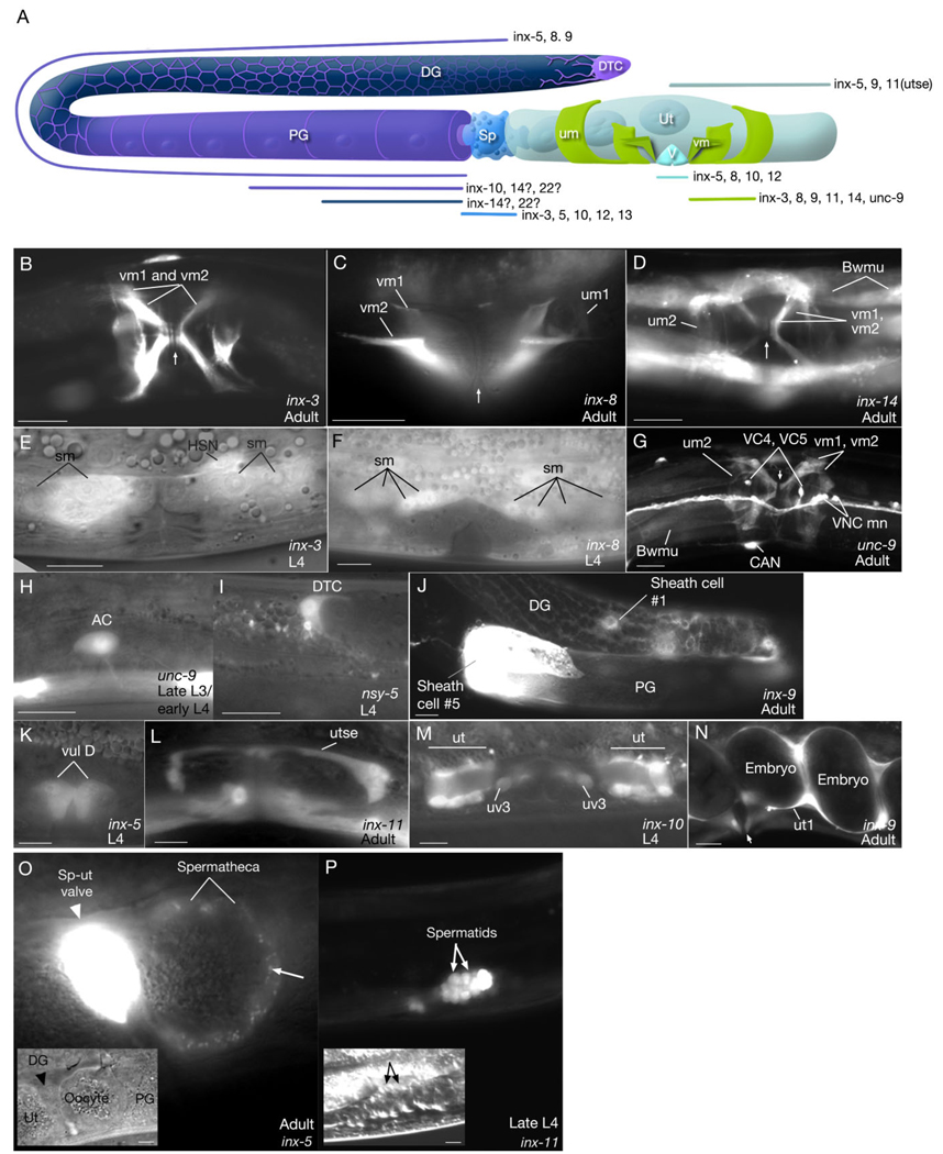
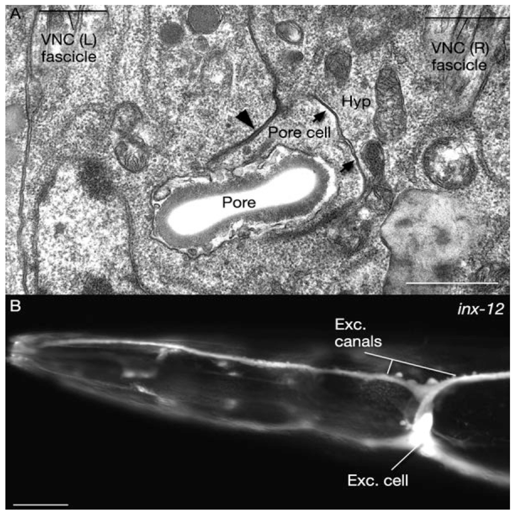
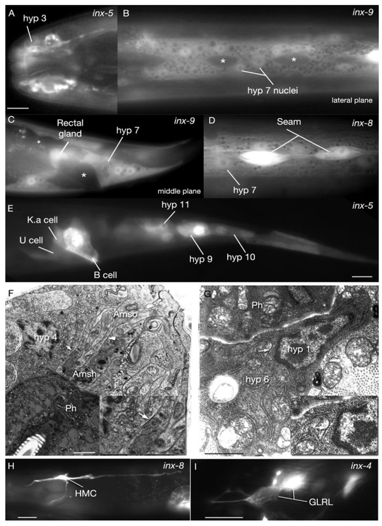
The innexin family of gap junction proteins has 25 members in Caenorhabditis elegans. Here, we describe the first high-resolution expression map of all members through analysis of live worms transformed with green fluorescent protein under the control of entire promoter regions. Our analyses show that innexins have dynamic expression patterns throughout development and are found in virtually all cell types and tissues. Complex tissues, such as the pharynx, intestine, gonad, as well as scaffolding tissues and guidepost cells express a variety of innexins in overlapping or complementary patterns, suggesting they may form heteromeric and heterotypic channels. Innexin expression occurs in several types of cells that are not known to form gap junctions as well as in a pair of migrating cells, suggesting they may have hemichannel function. Therefore, innexins likely play roles in almost all body functions, including embryonic development, cell fate determination, oogenesis, egg laying, pharyngeal pumping, excretion, and locomotion.


Figures

Fig. 1
Fig. 1
A: Graphic rendition of subunit composition of homotypic, heterotypic, and heteromeric gap junctions. B: Transcriptional fusion constructs used in this study. For all innexin genes except inx-21 and inx-22, a genomic DNA fragment containing a short upstream regulatory sequence of each innexin gene (black straight bars) was subcloned into a green fluorescent protein (GFP) expression vector which was then coinjected with the corresponding cosmid (dotted lines) into hermaphrodites allowing for in vivo homologous recombination. Because there were no suitable cosmids for inx-21 and inx-22, two long fragments upstream of the initiation sites of these two genes were polymerase chain reaction (PCR)-cloned and used for subcloning. T16H5 only contains the 706-bp sequence upstream of the nsy-5 initiation site, which might have revealed activating sequences in two neurons (AVK and AVB), which were not observed to express nsy-5 previously (Chuang et al., 2007). The transcription products of Punc-7a∷GFP and Punc-7b∷GFP are referred to as unc-7a and unc-7b, respectively, throughout this study.
Fig. 2
Fig. 2. Innexins in the alimentary canal
A: Graphic representation of expression domains of innexins in epithelial and muscle tissue is shown as black bars above the hermaphrodite image. Darker bars represent stronger adult expression. Thus, inx-11 is expressed strongly in the posterior intestine and faintly in the rest of the intestine. inx-21 is expressed solely in the intestine (light pink) in the adult, however the expression level is low. The numbers shown on the bi-lobed pharynx (green) indicate the pharyngeal muscles. A, arcades (light purple); pe, pharyngeal epithelium; vpi, pharyngeal valve (brown); vir, intestinal rectal valve (dark brown); rect epi, rectal epithelium (light brown). B: Fluorescent micrographs of transgenic animals expressing p inx∷GFP reporters. Left two columns show pharyngeal images. M3, M3 motor neuron; mc2, marginal cell 2; HMC, head mesodermal cell. Third column top panel shows g2 gland that expresses inx-3. Middle panel shows inx-15 expression in intestine and bottom panel shows inx-11 expression in intestinal rectal valve and also the posterior intestine (arrow). Rectal epithelial (K/K′ and B) innexin expressions are shown on the rightmost column. DVC, DVC neuron. C: Fluorescent micrographs of pinx-2∷GFP expression through developmental stages. i: Approximately 30-cell stage. inx-2 is expressed throughout the embryo. ii: Approximately embryonic day (E) 16 stage. Expression of inx-2 is stronger in the intestinal precursors (arrowhead) than the rest of the embryo although it continues to be expressed outside the alimentary canal. iii: Comma stage embryo. inx-2 expression is restricted to intestinal cells (arrowhead) and some pharyngeal precursors (arrow). iv: Three-fold stage. inx-2 expression continues at high levels in the intestinal cells (arrowhead) and terminal bulb pharyngeal muscle (arrow). v: L1 larva. inx-2 expression is decreased in the intestinal cells (arrowheads) although it continues to be expressed at relatively high levels in the pharyngeal muscle (arrow) and the pharyngeal–intestinal valve (middle inset, arrow). Scale bars = 10 µm.
Fig. 3
Fig. 3. Expression of innexins in reproductive tissues
A: Graphic representation of expression domains of innexins in the reproductive system (after Hall and Altun, 2008). DTC, distal tip cell (violet); DG, distal gonad; PG, proximal gonad; Sp, spermatheca (bright blue); um, uterine muscle (green); vm, vulva muscle (green); Ut, uterus (pale blue); V, vulva (sky blue). Gonad sheath is shown in purple color and germline in navy blue. inx-5, inx-8, inx-9 are expressed throughout gonad sheath (including thin fingers covering distal gonad) while inx-10 expression is higher in proximal gonad. inx-14 and inx-22 may be localized to oocytes and/or proximal gonad sheath (Whitten and Miller, 2007). B–D,G,J,N: Fluorescent micrographs of transgenic animals expressing the pinx∷GFP reporter genes. E,F,H,I,K–M: Fluorescent micrographs transposed over differential interference contrast microscopy (DIC) images of the same anatomical region. Scale bars = 10 µm. (B,D,G) ventral views; (C,E,F,H,I–N) lateral views. AC, anchor cell; Bwmu, body wall muscles; DG, distal gonad; DTC, distal tip cell; PG, proximal gonad; sm, sex muscle progenitors; um, uterine muscles; ut, uv, utse, portions of uterus; vm, vulval muscles; vul, vulval epithelium; VNCmn, ventral nerve cord motor neurons. Vulva position is marked with an arrow in some panels (B,C,D,F,N). The developmental stages of the animals are indicated on each panel. O,P: Innexin expression in the spermatheca and during spermatogenesis. O: Fluorescent micrograph of pinx-5∷GFP reporter gene expression in spermatheca superimposed to the DIC image (inset) of the same region in an adult hermaphrodite. Strong expression is observed in spermathecal-uterine (sp-ut) valve. An oocyte is seen inside the spermatheca. inx-5 is seen as localized to small punctate regions on the spermathecal wall (arrow), suggesting membrane localization signal is within the 30 amino acid sequence from the N-terminus included in our expression construct. Scale bar = 1 µm. Similar punctate/aggregated localization patterns were previously observed for inx-3 and inx-6 (Starich et al., 1996; Li et al., 2003). P: Fluorescent micrograph of pinx-11∷GFP reporter gene expression in the spermatids in a late L4 stage larva. (Inset) DIC image of the same anatomical region. Scale bar = 10 µm in inset.
Fig. 4
Fig. 4. Gap junctions and innexin expression in the excretory system
A: Horizontal section. Transmission electron micrograph of gap junctions (arrows) between the pore cell and the hypodermis. Pore cell also makes adherens junctions (arrowhead) to the hypodermis. Exc, excretory; Hyp, hypodermis; VNC, ventral nerve cord. Animal name: Hall/N533 (print number 4216). B: Fluorescent micrograph of expression of pinx-12∷GFP reporter gene in the excretory cell and its canals. Scale bars = 1 µm in A, 10 µm in B.
Fig. 5
Fig. 5. Innexins in the hypodermal (hyp) epithelium and mesoderm (all adult images)
Fluorescent micrographs of transgenic animals expressing the pinx∷GFP reporter genes; all lateral views, anterior to the left. A: pinx-5∷GFP is expressed in the anterior hypodermis. B,C: pinx-9∷GFP is expressed in the main body hypodermis (hyp 7) and rectal gland, but excluded from the seam (asterisks in B) and the rectal epithelium (asterisk in C). D: inx-8 is coexpressed in the seam and hypodermis. E: pinx-5∷GFP is expressed in the rectal epithelium (B, K.a, and U cells) and the posterior hypodermis (hyp 9–11). F,G: Gap junctions between glia and hypodermis and between hypodermal cells in the adult head. Transmission electron micrographs. Ph, pharynx. F: Gap junctions are seen between amphid socket (Amso) and amphid sheath (Amsh) glia (arrowhead) as well as the amphid sheath and hypodermis (hyp 4 cell; arrow). Lateral section; high pressure frozen material. G: A gap junction (arrow) is seen between hyp 1 and hyp 6 cells. (Note by this stage hyp 6 has fused to hyp 7; not shown.) Transverse section; immersion fixed material (MRC/N2T). Insets show some junctional regions at higher power. H,I: Innexin expression in two mesodermal tissues. Lateral views, anterior to the left. H. pinx-8∷GFP reporter gene expression in HMC (head mesodermal cell). I. pinx-4∷GFP reporter gene expression in GLRL. Scale bars = 5 µm in A, 10 µm in E (applies to B–E), 1 µm in F,G, 10 µm in H,I.

References

    1. Albertson DG, Thomson JN. The pharynx of Caenorhabditis elegans. Philos Trans R Soc Lond B Biol Sci. 1976;275:299–325. - PubMed
    1. Aurelio O, Hall DH, Hobert O. Immunoglobulin-domain proteins required for maintenance of ventral nerve cord organization. Science. 2002;295:686–690. - PubMed
    1. Avery L. The genetics of feeding in Caenorhabditis elegans. Genetics. 1993;133:897–917. - PMC - PubMed
    1. Avery L, Horvitz HR. Pharyngeal pumping continues after laser killing of the pharyngeal nervous system of C. elegans. Neuron. 1989;3:473–485. - PubMed
    1. Barbe MT, Monyer H, Bruzzone R. Cell-cell communication beyond connexins: the pannexin channels. Physiology (Bethesda) 2006;21:103–114. - PubMed

Publication types

MeSH terms

LinkOut - more resources