

Test-retest reproducibility of the default-mode network in healthy individuals
- PMID: 19621371
- PMCID: PMC6871144
- DOI: 10.1002/hbm.20860
Test-retest reproducibility of the default-mode network in healthy individuals
Abstract
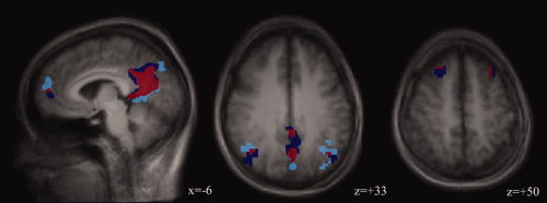
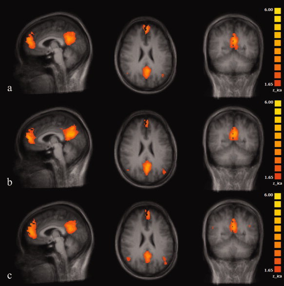
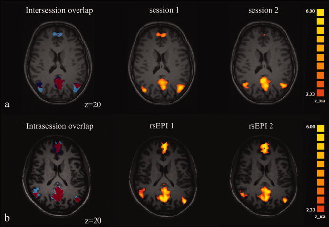
Independent component analysis (ICA) of functional magnetic resonance imaging (fMRI) time-series reveals distinct coactivation patterns in the resting brain representing spatially coherent spontaneous fluctuations of the fMRI signal. Among these patterns, the so-called default-mode network (DMN) has been attributed to the ongoing mental activity of the brain during wakeful resting state. Studies suggest that many neuropsychiatric diseases disconnect brain areas belonging to the DMN. The potential use of the DMN as functional imaging marker for individuals at risk for these diseases, however, requires that the components of the DMN are reproducible over time in healthy individuals. In this study, we assessed the reproducibility of the DMN components within and between imaging sessions in 18 healthy young subjects (mean age, 27.5 years) who were scanned three times with two resting state scans during each session at 3.0 T field strength. Statistical analysis of fMRI time-series was done using ICA implemented with BrainVoyager QX. At all three sessions the essential components of the DMN could be identified in each individual. Spatial extent of DMN activity and size of overlap within and between sessions were most reproducible for the anterior and posterior cingulate gyrus. The degree of reproducibility of the DMN agrees with the degree of reproducibility found with motor paradigms. We conclude that DMN coactivation patterns are reproducible in healthy young subjects. Therefore, these data can serve as basis to further explore the effects of aging and neuropsychiatric diseases on the DMN of the brain.
(c) 2009 Wiley-Liss, Inc.
Figures
References
-
- Alexander GE, Chen K, Pietrini P, Rapoport SI, Reiman EM ( 2002): Longitudinal PET evaluation of cerebral metabolic decline in dementia: A potential outcome measure in Alzheimer's disease treatment studies. Am J Psychiatry 159: 738–745. - PubMed
-
- Biswal B, Yetkin FZ, Haughton VM, Hyde JS ( 1995): Functional connectivity in the motor cortex of resting human brain using echo‐planar MRI. Magn Reson Med 34: 537–541. - PubMed
-
- D'Argembeau A, Collette F, Van der Linden M, Laureys S, Del Fiore G, Degueldre C, Luxen A, Salmon E ( 2005): Self‐referential reflective activity and its relationship with rest: A PET study. Neuroimage 25: 616–624. - PubMed
MeSH terms
LinkOut - more resources
Full Text Sources
Medical
Miscellaneous
