Review
. 2009 Sep-Oct;44(5):245-63.
doi: 10.1080/10409230903061207.

An intracellular trafficking pathway in the seminiferous epithelium regulating spermatogenesis: a biochemical and molecular perspective

Affiliations
Review

An intracellular trafficking pathway in the seminiferous epithelium regulating spermatogenesis: a biochemical and molecular perspective

C Yan Cheng et al. Crit Rev Biochem Mol Biol. 2009 Sep-Oct.

Abstract

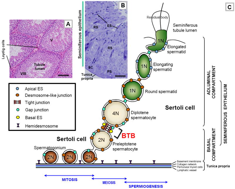
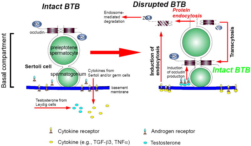
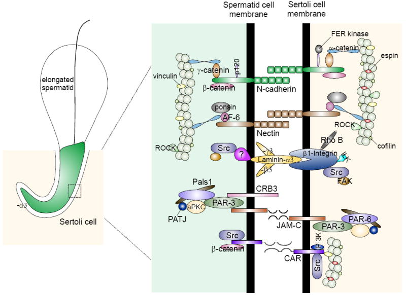
During spermatogenesis in adult rat testes, fully developed spermatids (i.e. spermatozoa) at the luminal edge of the seminiferous epithelium undergo "spermiation" at stage VIII of the seminiferous epithelial cycle. This is manifested by the disruption of the apical ectoplasmic specialization (apical ES) so that spermatozoa can enter the tubule lumen and to complete their maturation in the epididymis. At the same time, the blood-testis barrier (BTB) located near the basement membrane undergoes extensive restructuring to allow transit of preleptotene spermatocytes so that post-meiotic germ cells complete their development behind the BTB. While spermiation and BTB restructuring take place concurrently at opposite ends of the Sertoli cell epithelium, the biochemical mechanism(s) by which they are coordinated were not known until recently. Studies have shown that fragments of laminin chains are generated from the laminin/integrin protein complex at the apical ES via the action of MMP-2 (matrix metalloprotease-2) at spermiation. These peptides serve as the local autocrine factors to destabilize the BTB. These laminin peptides also exert their effects on hemidesmosome which, in turn, further potentiates BTB restructuring. Thus, a novel apical ES-BTB-hemidesmosome regulatory loop is operating in the seminiferous epithelium to coordinate these two crucial cellular events of spermatogenesis. This functional loop is further assisted by the Par3/Par6-based polarity protein complex in coordination with cytokines and testosterone at the BTB. Herein, we provide a critical review based on the latest findings in the field regarding the regulation of these cellular events. These recent findings also open up a new window for investigators studying blood-tissue barriers.


Figures

Figure 1
Figure 1. The general morphological features of mammalian testes, and a schematic drawing of the seminiferous epithelium illustrating the relative location of different junction types, and the adluminal (apical) and basal compartments of the seminiferous epithelium created by the blood-testis barrier (BTB) in adult rat testes
(A) This micrograph is the cross-section of an adult rat testis, showing several seminiferous tubules (e.g., a stage V and a stage VIII seminiferous tubule), which are the functional units that produce spermatozoa. The sperm producing function of the seminiferous tubule during spermatogenesis is also supported by testosterone produced by Leydig cells which are found in the interstitium. Bar = 80 μm. (B) This is the magnified view of a stage V tubule, illustrating the different germ cell types (e.g., PS, pachytene spermatocyte; RS, round spermatid; ES, elongating spermatid) are tightly associated with Sertoli cells (SC, Sertoli cell nucleus) found in the seminiferous epithelium. Bar = 8 μm. (C) This is the schematic drawing illustrating the seminiferous epithelium is composed of Sertoli cells and germ cells at different stages of their development, lying on the tunica propria which is composed of two acellular zones: basement membrane (a modified form of extracellular matrix) and type I collagen network; and two cellular zones: peritubular myoid cells and lymphatic vessel. The BTB, located near the basement membrane, is constituted by coexisting tight junction, gap junction, desmosome-like junction, and basal ectoplasmic specialization (basal ES), between adjacent Sertoli cells. The BTB also physically divides the seminiferous epithelium into the basal and adluminal (apical) compartments and confers cell polarity in the seminiferous epithelium. From step 8 through step 19 spermatids, apical ectoplasmic specialization (apical ES) is the only anchoring device that maintains cell adhesion and orientation of a developing spermatid onto the Sertoli cell in the seminiferous epithelium. Preleptotene spermatocytes differentiated from type B spermatogonia are the germ cells in transit at the BTB at stage VIII of the seminiferous epithelial cycle while differentiating into leptotene and zygotene spermatocytes. Once these primary spermatocytes enter the adluminal compartment, they differentiate into pachytene spermatocytes and diplotene spermatocytes to begin meiosis I to be followed immediately by meiosis II to form haploid round spermatids at stage XIV of the epithelial cycle in rats. Round spermatids undergo spermiogenesis via steps 1 through step 19 spermatids in rats, which is typified by the condensation of the genetic materials. The genetic substances are tightly packed into the spermatid head concomitant with the formation of the acrosome above the head region and the elongation of the spermatid tail. During these morphological changes, developing spermatids are also in transit near the BTB towards the luminal edge of the seminiferous epithelium progressively, then migrate towards the basement membrane (such as at stage IV-V of the epithelial cycle) (see Fig. 1B) and moving towards the luminal edge again (such as stage VI-VII of the epithelial cycle) until the fully developed spermatids (i.e., spermatozoa) are emptied into the seminiferous tubule lumen at spermiation at stage VIII of the epithelial cycle which is associated with disruption of the adhesion protein complexes at the elongated spermatid-Sertoli cell interface, and the shedding of the residual bodies. The Sertoli cell also serves as a scavenger by engulfing the residual bodies via phagocytosis. As a result of this germ cell maturation process and the continuous movement of developing germ cells, in particular elongating spermatids, across the seminiferous epithelium, there are extensive restructuring, namely disassembly and reassembly, of junctions at the Sertoli-Sertoli and Sertoli-germ cell interface during spermatogenesis. Also noted are the three phases of spermatogenesis: mitosis, meiosis, and spermiogenesis (see text for details). 1N, haploid; 2N, diploid; 4N, tetraploid.
Figure 2
Figure 2. The protein complexes and their peripheral regulators and adaptors that constitute the BTB in the rat testis
As described in the text that the BTB in the testis is constituted by integral membrane proteins of the: (i) tight junction (TJ): occludins, claudins, tricellulin, JAMs, and CAR; (ii) desmosome-like junction: desmogleins, desmocollins; (iii) gap junction (GJ): connexins; and (iv) basal ES: N-cadherins, nectins. Each integral membrane protein is associated with its corresponding adaptors (e.g., ZOs, plakophilins, catenins, afadins), regulatory proteins (e.g., FAK, MAPKs, ILK, FER kinase), polarity proteins (e.g., Par, Pals1). All these protein complexes, in turn, attach to actin via actin-binding proteins (e.g., vinculin, afadin, espin) except for desmosome-like junction which uses intermediate filaments (e.g., vimentin) for attachment.
Figure 3
Figure 3. A hypothetical model illustrating the likely mechanism utilized by the testis to facilitate the transit of primary preleptotene spermatocytes at the BTB while maintaining the immunological barrier function
At the time of primary preleptotene spermatocytes in transit at the BTB, such as at stage VIII of the epithelial cycle, testosterone produced by Leydig cells in the interstitium enters the BTB microenvironment, which is known to promote BTB function, binds to androgen receptor to exert is effects or mediates its action via nongenomic pathway, such as c-Src and ERK (Cheng et al., 2007; Fix et al., 2004) (see left panel). This promotes the production of BTB proteins (e.g., claudin-11, occludin) (Kaitu’u-Lino et al., 2007; Yan et al., 2008c) for the assembling of ‘new’ TJ-fibrils behind a migrating primary preleptotene spermatocyte (see right panel). Furthermore, testosterone also appears to promote transcytosis of internalized integral membrane proteins, perhaps from the apical region to the basal region of the migrating spermatocytes to facilitate ‘new’ TJ-fibrils assembly (Yan et al., 2008c). Cytokines (e.g., TGF-β3, TNFα) released from Sertoli and germ cells also bind to their corresponding receptors at the BTB to activate the downstream signaling function, which, in turn, induces protein endocytosis at the ‘old’ BTB site above the migrating spermatocytes but targeted to degradation via the endosome-mediated pathway (Yan et al., 2008c) (left panel). Thus, ‘new’ TJ-fibrils behind the migrating spermatocytes continue to maintain the immunological barrier while the ‘old’ TJ-fibrils above the spermatocytes in transit are disrupted to facilitate cell movement. This thus provides a novel mechanism to maintain the immunological barrier during the transit of primary preleptotene spermatocytes at the BTB.
Figure 4
Figure 4. The protein complexes and their peripheral regulators and adaptors that constitute the apical ES in the rat testis
The presently known integral membrane proteins at the apical ES in adult rat testes are: cadherins, nectins, integrins, laminins/receptor protein(s) (note: laminins, such as α3, β3 and γ3 are cell surface proteins without any transmembrane domain, residing on elongating/elongated spermatids that anchor to spermatids at the apical ES via a yet-to-be identified laminin receptor protein), CRB3, JAM-C, and CAR. Each of these protein binds to its corresponding adaptors (e.g., catenins, afadins), polarity proteins (e.g., Par, Pals1), and protein kinases (e.g., c-Src, FAK), using actin for its attachment. Apical ES is an atypical adherens junction (AJ) type (Wong et al., 2008a) since it is composed of proteins usually restricted to cell-matrix focal adhesion complex (or focal contact) (e.g., c-Src, FAK), and TJ (e.g., JAM-C, CAR).
Figure 5
Figure 5. A schematic drawing illustrating the presence of a local regulatory loop known as the “apical ES-BTB-hemidesmosome” in the seminiferous epithelium to coordinate the events of spermiation and BTB restructuring that facilitate the transit of primary preleptotene spermatocytes at stage VIII of the seminiferous epithelial cycle
The left panel depicts the seminiferous epithelium of a normal rat testis at stage VII of the epithelial cycle. As described in the text, at the time of spermiation (see right panel), MMP-2 cleaves laminin-333 to release biologically active fragments at the time of apical ES disruption (Step i), which can either destabilize BTB (Step ii) directly by reducing the steady-state level of integral membrane proteins at the site via protein endocytosis and endosome-mediated degradation, or indirectly by perturbing the hemidesmosome function by reducing the steady-state level of β1-integrin. The disruption of the hemidesmosome (Step iii), in turn, perturbs the BTB integrity via a mechanism remains to be investigated. However, it likely involves FAK and/or cSrc. The endocytosed proteins may also be re-used to assemble ‘new ’ TJ-fibrils behind the primary spermatocyte in transit via transcytosis. Using such a mechanism, the events of spermiation and BTB restructuring that take place at the opposite ends of the seminiferous epithelium can be coordinated at stage VIII of the epithelial cycle.

Similar articles

Cited by

References

    1. Al-Sadi R, Boivin M, T M. Mechanism of cytokine modulation of epithelial tight junction barrier. Front Biosci. 2009;14:2765–2778. - PMC - PubMed
    1. Alberts B, Johnson A, Lewis J, Raff M, Roberts K, Walter P. Molecular Biology of the Cell. New York: Garland Science; 2002.
    1. Aumailley M, Bruckner-Tuderman L, Carter W, Deutzmann R, Edgar D, Ekblom P, Engel J, Engvall E, Hohenester E, Jones J, et al. A simplified laminin nomenclature. Matrix Biol. 2005;24:326–332. - PubMed
    1. Bart J, Hollema H, Groen HJ, de Vries EG, Hendrikse NH, Sleijfer DT, Wegman TD, Vaalburg W, van der Graaf WT. The distribution of drug-efflux pumps, P-gp, BCRP, MRP1 and MRP2, in the normal blood-testis barrier and in primary testicular tumours. Eur J Cancer. 2004;40:2064–2070. - PubMed
    1. Bernacki J, Dobrowolska A, Nierwinska K, Malecki A. Physiology and pharmacological role of the blood-brain barrier. Pharmacol Rep. 2008;60:600–622. - PubMed

Publication types

LinkOut - more resources