

Invasiveness as a putative additional virulence mechanism of some atypical Enteropathogenic Escherichia coli strains with different uncommon intimin types
- PMID: 19622141
- PMCID: PMC2724384
- DOI: 10.1186/1471-2180-9-146
Invasiveness as a putative additional virulence mechanism of some atypical Enteropathogenic Escherichia coli strains with different uncommon intimin types
Erratum in
- BMC Microbiol. 2009;9:235
Abstract
Background: Enteropathogenic Escherichia coli (EPEC) produce attaching/effacing (A/E) lesions on eukaryotic cells mediated by the outer membrane adhesin intimin. EPEC are sub-grouped into typical (tEPEC) and atypical (aEPEC). We have recently demonstrated that aEPEC strain 1551-2 (serotype O non-typable, non-motile) invades HeLa cells by a process dependent on the expression of intimin sub-type omicron. In this study, we evaluated whether aEPEC strains expressing other intimin sub-types are also invasive using the quantitative gentamicin protection assay. We also evaluated whether aEPEC invade differentiated intestinal T84 cells.
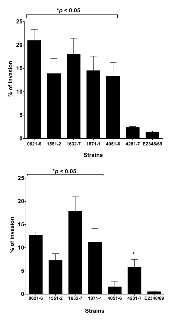
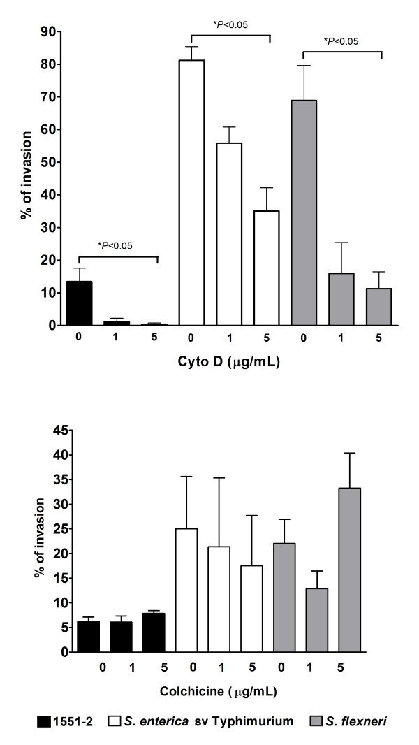
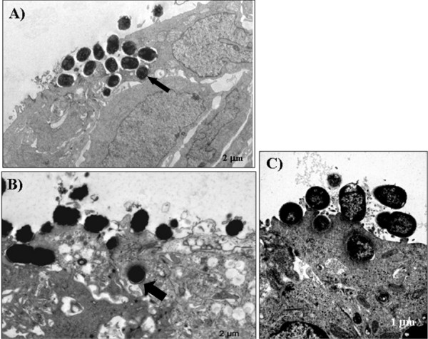
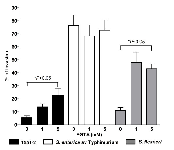
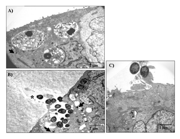
Results: Five of six strains invaded HeLa and T84 cells in a range of 13.3%-20.9% and 5.8%-17.8%, respectively, of the total cell-associated bacteria. The strains studied were significantly more invasive than prototype tEPEC strain E2348/69 (1.4% and 0.5% in HeLa and T84 cells, respectively). Invasiveness was confirmed by transmission electron microscopy. We also showed that invasion of HeLa cells by aEPEC 1551-2 depended on actin filaments, but not on microtubules. In addition, disruption of tight junctions enhanced its invasion efficiency in T84 cells, suggesting preferential invasion via a non-differentiated surface.
Conclusion: Some aEPEC strains may invade intestinal cells in vitro with varying efficiencies and independently of the intimin sub-type.
Figures
Similar articles
-
Invasion of differentiated intestinal Caco-2 cells is a sporadic property among atypical enteropathogenic Escherichia coli strains carrying common intimin subtypes.Pathog Dis. 2014 Mar;70(2):167-75. doi: 10.1111/2049-632X.12112. Epub 2013 Dec 16. Pathog Dis. 2014. PMID: 24339197
-
The localized adherence pattern of an atypical enteropathogenic Escherichia coli is mediated by intimin omicron and unexpectedly promotes HeLa cell invasion.Cell Microbiol. 2008 Feb;10(2):415-25. doi: 10.1111/j.1462-5822.2007.01054.x. Epub 2007 Oct 2. Cell Microbiol. 2008. PMID: 17910741
-
Expression of the locus of enterocyte effacement genes during the invasion process of the atypical enteropathogenic Escherichia coli 1711-4 strain of serotype O51:H40.Microbiol Spectr. 2024 Oct 3;12(10):e0030424. doi: 10.1128/spectrum.00304-24. Epub 2024 Aug 27. Microbiol Spectr. 2024. PMID: 39189752 Free PMC article.
-
An overview of atypical enteropathogenic Escherichia coli.FEMS Microbiol Lett. 2009 Aug;297(2):137-49. doi: 10.1111/j.1574-6968.2009.01664.x. Epub 2009 May 27. FEMS Microbiol Lett. 2009. PMID: 19527295 Review.
-
Enteropathogenic Escherichia coli: foe or innocent bystander?Clin Microbiol Infect. 2015 Aug;21(8):729-34. doi: 10.1016/j.cmi.2015.01.015. Epub 2015 Jan 28. Clin Microbiol Infect. 2015. PMID: 25726041 Free PMC article. Review.
Cited by
-
Genetic evaluation of Locus of enterocyte effacement pathogenicity island (LEE) in Enteropathogenic Escherichia coli isolates (EPEC).Iran J Microbiol. 2013 Dec;5(4):345-9. Iran J Microbiol. 2013. PMID: 25848503 Free PMC article.
-
Escherichia albertii Pathogenesis.EcoSal Plus. 2020 Jun;9(1):10.1128/ecosalplus.ESP-0015-2019. doi: 10.1128/ecosalplus.ESP-0015-2019. EcoSal Plus. 2020. PMID: 32588811 Free PMC article. Review.
-
Two atypical enteropathogenic Escherichia coli strains induce the production of secreted and membrane-bound mucins to benefit their own growth at the apical surface of human mucin-secreting intestinal HT29-MTX cells.Infect Immun. 2010 Mar;78(3):927-38. doi: 10.1128/IAI.01115-09. Epub 2010 Jan 11. Infect Immun. 2010. PMID: 20065027 Free PMC article.
-
Microbiology and Epidemiology of Escherichia albertii-An Emerging Elusive Foodborne Pathogen.Microorganisms. 2022 Apr 22;10(5):875. doi: 10.3390/microorganisms10050875. Microorganisms. 2022. PMID: 35630320 Free PMC article. Review.
-
The transmembrane domains of the type III secretion system effector Tir are involved in its secretion and cellular activities.Front Cell Infect Microbiol. 2023 Feb 14;13:1103552. doi: 10.3389/fcimb.2023.1103552. eCollection 2023. Front Cell Infect Microbiol. 2023. PMID: 36864885 Free PMC article.
References
-
- Kaper JB. Defining EPEC. Rev Microbiol. 1996;27:130–133.
Publication types
MeSH terms
Substances
LinkOut - more resources
Full Text Sources
