

Establishment and characterization of pleomorphic adenoma cell systems: an in-vitro demonstration of carcinomas arising secondarily from adenomas in the salivary gland
- PMID: 19622142
- PMCID: PMC2722671
- DOI: 10.1186/1471-2407-9-247
Establishment and characterization of pleomorphic adenoma cell systems: an in-vitro demonstration of carcinomas arising secondarily from adenomas in the salivary gland
Abstract
Background: Among the salivary gland carcinomas, carcinoma in pleomorphic adenoma has been regarded as a representative carcinoma type which arises secondarily in the background of a pre-existent benign pleomorphic adenoma. It is still poorly understood how and which benign pleomorphic adenoma cells transform into its malignant form, carcinoma ex pleomorphic adenoma.
Methods: We have established five cell systems from a benign pleomorphic adenoma of the parotid gland of a 61-year-old woman. They were characterized by immunofluorescence, classical cytogenetics, p53 gene mutational analysis, fluorescence in-situ hybridization, and histopathological and immunohistochemical examinations of their xenografts, to demonstrate their potency of secondary transformation.
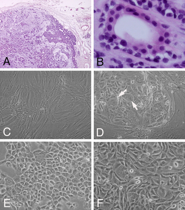
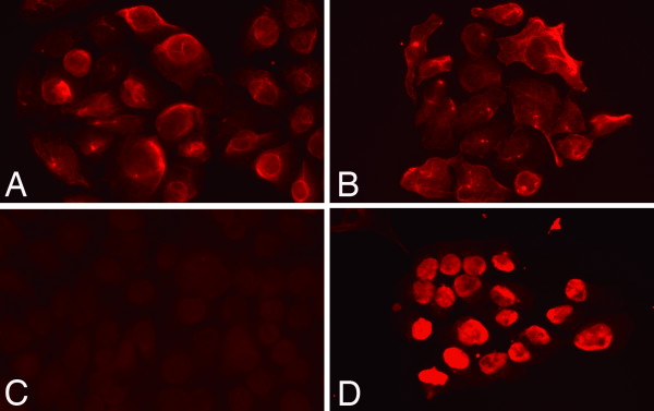
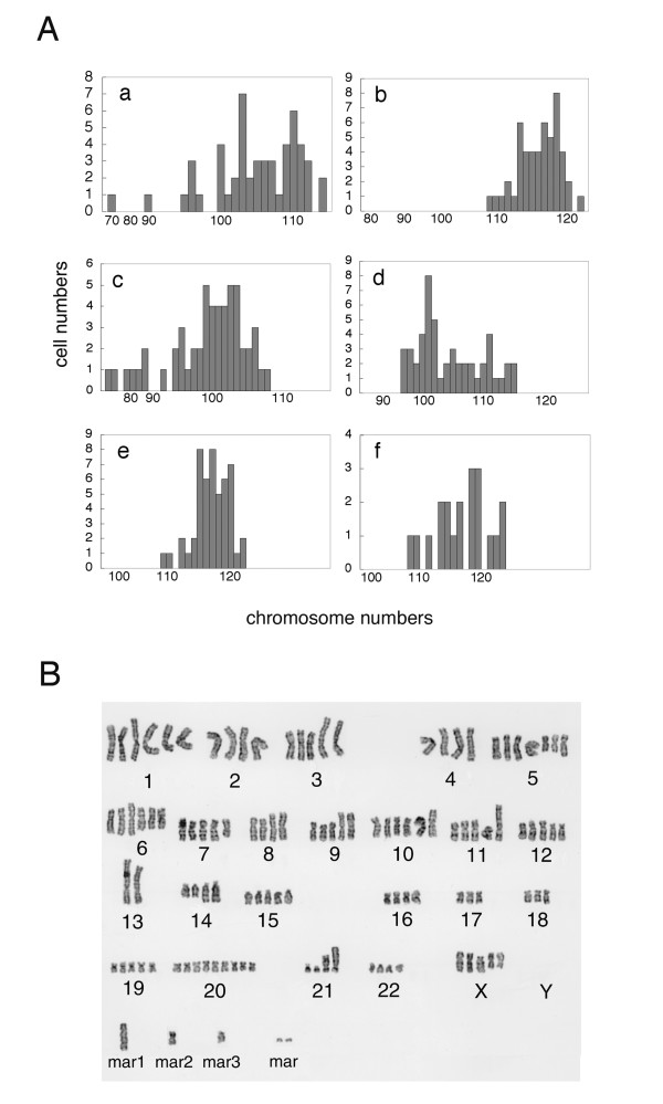
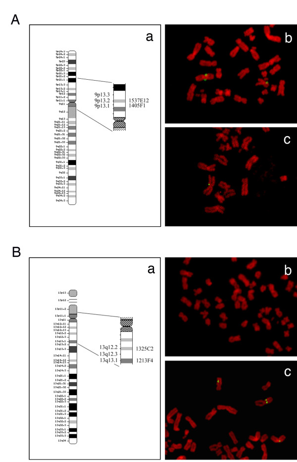
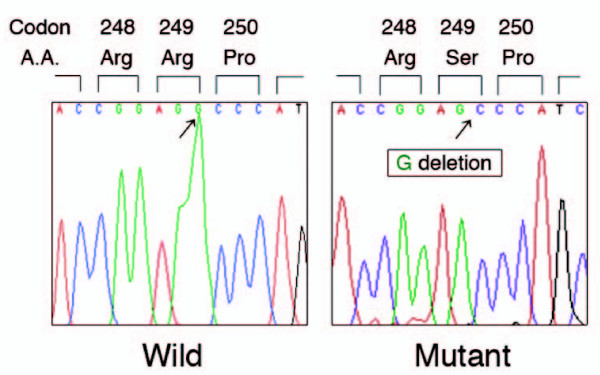
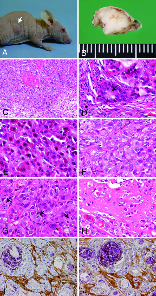
Results: We established and characterized five cell systems (designated as SM-AP1 to SM-AP5) from a benign pleomorphic adenoma of the parotid gland. SM-AP1 to SM-AP3 showed polygonal cell shapes while SM-AP4 and SM-AP5 were spindle-shaped. SM-AP1-3 cells were immunopositive for keratin only, indicating their duct-epithelial or squamous cell differentiation, while SM-AP4/5 cells were positive for both keratin and S-100 protein, indicating their myoepithelial cell differentiation. Chromosome analyses showed numeral abnormalities such as 5n ploidies and various kinds of structural abnormalities, such as deletions, translocations, derivatives and isodicentric chromosomes. Among them, der(9)t(9;13)(p13.3;q12.3) was shared by all five of the cell systems. In addition, they all had a common deletion of the last base G of codon 249 (AGG to AG_) of the p53 gene, which resulted in generation of its nonsense gene product. Transplanted cells in nude mice formed subcutaneous tumors, which had histological features of squamous cell carcinoma with apparent keratinizing tendencies. In addition, they had ductal arrangements or plasmacytoid appearances of tumor cells and myxoid or hyaline stromata, indicating some characteristics of pleomorphic adenoma.
Conclusion: This study demonstrates in vitro that certain cell types from pleomorphic adenoma are able to clone and survive over a long term and develop subcutaneous tumors in nude mice. The histological features of squamous cell carcinoma from the transplanted cell systems in nude mice might suggest a secondary onset of malignancy from a pre-existing benign adenoma.
Figures
Similar articles
-
PLAG1 gene alterations in salivary gland pleomorphic adenoma and carcinoma ex-pleomorphic adenoma: a combined study using chromosome banding, in situ hybridization and immunocytochemistry.Mod Pathol. 2005 Aug;18(8):1048-55. doi: 10.1038/modpathol.3800386. Mod Pathol. 2005. PMID: 15920557
-
Podoplanin is a novel myoepithelial cell marker in pleomorphic adenoma and other salivary gland tumors with myoepithelial differentiation.Virchows Arch. 2013 Mar;462(3):297-305. doi: 10.1007/s00428-012-1359-z. Epub 2012 Dec 22. Virchows Arch. 2013. PMID: 23262786
-
Cytogenetic analysis of salivary gland type tumors.Oral Surg Oral Med Oral Pathol Oral Radiol Endod. 1996 Aug;82(2):187-92. doi: 10.1016/s1079-2104(96)80223-x. Oral Surg Oral Med Oral Pathol Oral Radiol Endod. 1996. PMID: 8863309
-
The pleomorphic adenoma of salivary glands transplanted on athmymic mice. A lightmicroscopical and immunohistochemical investigation.Virchows Arch A Pathol Anat Histopathol. 1985;408(2-3):191-209. doi: 10.1007/BF00707982. Virchows Arch A Pathol Anat Histopathol. 1985. PMID: 2417405 Review.
-
[Pleomorphic adenoma of salivary glands: diagnostic pitfalls and mimickers of malignancy].Cesk Patol. 2012 Oct;48(4):179-83. Cesk Patol. 2012. PMID: 23121026 Review. Czech.
Cited by
-
A novel cell line derived from pleomorphic adenoma expresses MMP2, MMP9, TIMP1, TIMP2, and shows numeric chromosomal anomalies.PLoS One. 2014 Aug 19;9(8):e105231. doi: 10.1371/journal.pone.0105231. eCollection 2014. PLoS One. 2014. PMID: 25137137 Free PMC article.
-
Extracellular matrix remodelling and stiffening contributes to tumorigenesis of salivary carcinoma ex pleomorphic adenoma--A study based on patient-derived organoids.Cell Biosci. 2023 Jul 1;13(1):122. doi: 10.1186/s13578-023-01071-x. Cell Biosci. 2023. PMID: 37393249 Free PMC article.
-
GSK3β and CREB3 gene expression profiling in benign and malignant salivary gland tumors.Iran Biomed J. 2012;16(3):140-4. doi: 10.6091/IBJ.1050.2012. Iran Biomed J. 2012. PMID: 23023215 Free PMC article.
-
ECT2+ cell group acts as cancer stem cell in malignant pleomorphic adenoma.NPJ Precis Oncol. 2025 Jun 17;9(1):189. doi: 10.1038/s41698-025-00974-x. NPJ Precis Oncol. 2025. PMID: 40523903 Free PMC article.
-
Noninvasive carcinoma ex pleomorphic adenoma of the parotid gland: A difficult diagnosis on fine needle aspiration.Cytojournal. 2015 Apr 29;12:7. doi: 10.4103/1742-6413.156080. eCollection 2015. Cytojournal. 2015. PMID: 25972908 Free PMC article.
References
-
- Gnepp DR, Wening BM. In: Surgical pathology of the Salivary Glands. Ellis GL, Auclair PL, Gnepp DR, editor. Philadelphia: W.B. Saunders; 1991. Malignant mixed tumors; pp. 350–368.
-
- Ellis GL, Auclair PL. Tumor of the Salivary Glands: Atlas of Tumor Pathology, 3rd Series. Washington, DC: Armed Forces Institute of Pathology; 1996. pp. 228–238.
-
- Ohtaké S, Cheng J, Ida H, Suzuki M, Ohshiro K, Zhang W, Saku T. Precancerous foci in pleomorphic adenoma of the salivary gland: recognition of focal carcinoma and atypical tumor cells by P53 immunohistochemistry. J Oral Pathol Med. 2002;31:590–597. doi: 10.1034/j.1600-0714.2002.00040.x. - DOI - PubMed
-
- Brandwein M, Huvos AG, Dardick I, Thomas MJ, Theise ND. Noninvasive and minimally invasive carcinoma ex mixed tumor: A clinicopathologic and ploidy study of 12 patients with major salivary tumors of low (or no?) malignant potential. Oral Surg Oral Med Oral Pathol Radiol Endod. 1996;81:655–664. doi: 10.1016/S1079-2104(96)80071-0. - DOI - PubMed
-
- Auclair PL, Ellis GL. Atypical features in salivary gland mixed tumors: their relationship to malignant transformation. Mod Pathol. 1996;9:652–657. - PubMed
Publication types
MeSH terms
LinkOut - more resources
Full Text Sources
Medical
Research Materials
Miscellaneous
