. 2009 Jul 21:10:329.
doi: 10.1186/1471-2164-10-329.

Polymorphic segmental duplication in the nematode Caenorhabditis elegans

Affiliations

Polymorphic segmental duplication in the nematode Caenorhabditis elegans

Ismael A Vergara et al. BMC Genomics. .

Abstract

Background: The nematode Caenorhabditis elegans was the first multicellular organism to have its genome fully sequenced. Over the last 10 years since the original publication in 1998, the C. elegans genome has been scrutinized and the last gaps were filled in November 2002, which present a unique opportunity for examining genome-wide segmental duplications.

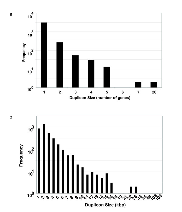
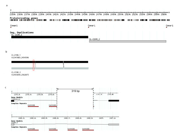
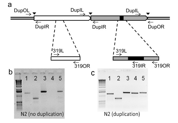
Results: Here, we performed analysis of the C. elegans genome in search for segmental duplications using a new tool -- OrthoCluster -- we have recently developed. We detected 3,484 duplicated segments -- duplicons -- ranging in size from 234 bp to 108 Kb. The largest pair of duplicons, 108 kb in length located on the left arm of Chromosome V, was further characterized. They are nearly identical at the DNA level (99.7% identity) and each duplicon contains 26 putative protein coding genes. Genotyping of 76 wild-type strains obtained from different labs in the C. elegans community revealed that not all strains contain this duplication. In fact, only 29 strains carry this large segmental duplication, suggesting a very recent duplication event in the C. elegans genome.

Conclusion: This report represents the first demonstration that the C. elegans laboratory wild-type N2 strains has acquired large-scale differences.


Figures

Figure 1
Figure 1
Size distribution of perfect duplicons in C. elegans genome. (a) Size distribution of all perfect duplicons in the C. elegans genome measured in number of genes. (b) Size distribution of all perfect duplicons in the C. elegans genome measured in kb. Each N value in the x-axis represent all those duplicons that fall in the range [N-1..N) kb. The y-axis represents the frequency in a logarithmic scale (base 10) of the frequency of a specific duplicon size. Thus, those bins with no visible bar mean that only one duplicon is observed for that particular value.
Figure 2
Figure 2
The two largest duplicons in the C. elegans genome. (a) Genome browser image of the largest duplicons CL-2198_1 (depicted in black) and CL-2198_2 (depicted in gray), and flanking Cemar1 transposons (shown in red). (b) Alignment of the two largest duplicons indicate the locations of the small differences. From 5' to 3': (1) 319 bp deletion in first duplicon. (2) A single nucleotide insertion ('C') in first duplicon at 2,381,150 bp. (3) A single nucleotide difference ('T" is the first duplicon at 2,420,123 bp and 'C' in the second duplicon at 2,528,402 bp) (4) A single nucleotide difference ('A' in the first duplicon at 2,420,126 bp and 'T' in the second duplicon at 2,528,405 bp). (5) A single nucleotide difference ('T' in the first duplicon at 2,420,132 bp and 'C' in second duplicon at 2,528,411 bp). (6) A triplet difference ('TAC' in the first duplicon from 2,420,134 bp to 2,420,136 bp and 'ACT' in the second duplicon from 2,528,413 bp to 2,528,415 bp). (c) The 319 bp unique sequence in the largest duplicon. Multiple copies of Ce000266 repetitive element are located in the region. The upper and lower panels show the upstream and downstream copies of the largest duplicons, respectively.
Figure 3
Figure 3
PCR analysis of the largest tandem segmental duplicons. (a) A schematic illustration of the largest duplicons, with PCR primers used for genotyping labeled. (b) A representative gel for strains that do not carry the largest duplication. (c) A representative gel for strains carrying the largest duplication. Lane 1 shows PCR product using primers 319L and 319OR; lane 2 shows PCR product using primers 319L and 319IR; lane 3 shows PCR product using primers DupOL and DupIR; lane 4 shows PCR product using primers DupIL and DupIR; and lane 5 shows PCR product using primers DupIL and DupOR.
Figure 4
Figure 4
Gene F56A4.3 at the junction of the largest pair of duplicons. F56A4.3 gene model (shown in the "Gene Models" track) is fully supported by an EST sequence (shown in the "ESTs aligned by BLAT (best)" track). The black and grey bars represent the ends of the largest pair of duplicons.

References

    1. Sturtevant AH. The Effects of Unequal Crossing over at the Bar Locus in Drosophila. Genetics. 1925;10(2):117–147. - PMC - PubMed
    1. Bailey JA, Eichler EE. Primate segmental duplications: crucibles of evolution, diversity and disease. Nat Rev Genet. 2006;7(7):552–564. doi: 10.1038/nrg1895. - DOI - PubMed
    1. Ohno S. Evolution by Gene Duplication. Berlin: Springer-Verlag; 1970.
    1. Troemel ER, Chou JH, Dwyer ND, Colbert HA, Bargmann CI. Divergent seven transmembrane receptors are candidate chemosensory receptors in C. elegans. Cell. 1995;83(2):207–218. doi: 10.1016/0092-8674(95)90162-0. - DOI - PubMed
    1. Robertson HM. Two large families of chemoreceptor genes in the nematodes Caenorhabditis elegans and Caenorhabditis briggsae reveal extensive gene duplication, diversification, movement, and intron loss. Genome Res. 1998;8(5):449–463. - PubMed

Publication types

Substances

LinkOut - more resources