. 2009 Jul 21:10:86.
doi: 10.1186/1471-2202-10-86.

c-MycERTAM transgene silencing in a genetically modified human neural stem cell line implanted into MCAo rodent brain

Affiliations

c-MycERTAM transgene silencing in a genetically modified human neural stem cell line implanted into MCAo rodent brain

Lara Stevanato et al. BMC Neurosci. .

Abstract

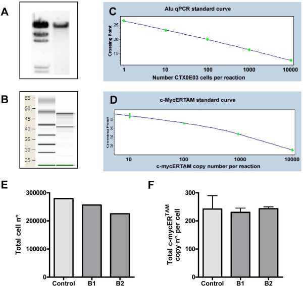
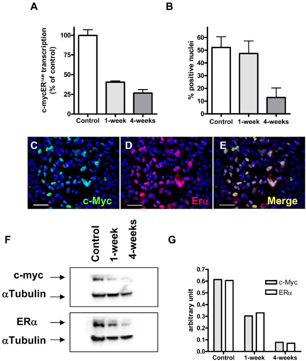
Background: The human neural stem cell line CTX0E03 was developed for the cell based treatment of chronic stroke disability. Derived from fetal cortical brain tissue, CTX0E03 is a clonal cell line that contains a single copy of the c-mycERTAM transgene delivered by retroviral infection. Under the conditional regulation by 4-hydroxytamoxifen (4-OHT), c-mycERTAM enabled large-scale stable banking of the CTX0E03 cells. In this study, we investigated the fate of this transgene following growth arrest (EGF, bFGF and 4-OHT withdrawal) in vitro and following intracerebral implantation into a mid-cerebral artery occluded (MCAo) rat brain. In vitro, 4-weeks after removing growth factors and 4-OHT from the culture medium, c-mycERTAM transgene transcription is reduced by ~75%. Furthermore, immunocytochemistry and western blotting demonstrated a concurrent decrease in the c-MycERTAM protein. To examine the transcription of the transgene in vivo, CTX0E03 cells (450,000) were implanted 4-weeks post MCAo lesion and analysed for human cell survival and c-mycERTAM transcription by qPCR and qRT-PCR, respectively.

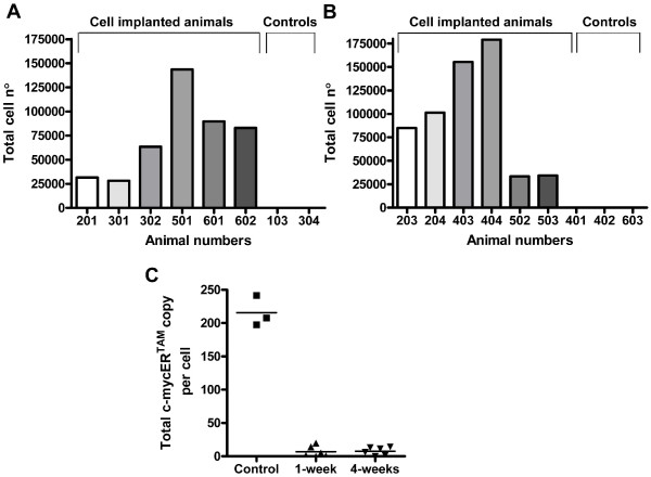
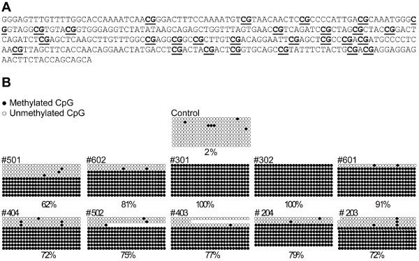
Results: The results show that CTX0E03 cells were present in all grafted animal brains ranging from 6.3% to 39.8% of the total cells injected. Prior to implantation, the CTX0E03 cell suspension contained 215.7 (SEM = 13.2) copies of the c-mycERTAM transcript per cell. After implantation the c-mycERTAM transcript copy number per CTX0E03 cell had reduced to 6.9 (SEM = 3.4) at 1-week and 7.7 (SEM = 2.5) at 4-weeks. Bisulfite genomic DNA sequencing of the in vivo samples confirmed c-mycERTAM silencing occurred through methylation of the transgene promoter sequence.

Conclusion: In conclusion the results confirm that CTX0E03 cells downregulated c-mycERTAM transgene expression both in vitro following EGF, bFGF and 4-OHT withdrawal and in vivo following implantation in MCAo rat brain. The silencing of the c-mycERTAM transgene in vivo provides an additional safety feature of CTX0E03 cells for potential clinical application.


Figures

Figure 1
Figure 1
Alu sequence and c-mycERTAM assay development and validation. Gels showing the quality and purity of gDNA by agarose gel electrophoresis (A) and RNA by virtual gel produced by Agilent 2100 Bioanalyzer (B, RNA Integrity Number >9.4 as analyzed by Agilent RNA 600 nano kit [32]) isolated from the same sample. Standard curves used to determine: cell number by Alu sequence qPCR (C, Error 0.0300, efficiency 1.993; 3 replicates); absolute c-mycERTAM copy number by c-mycERTAM qRT-PCR (D, Error 0.0837 and efficiency 2.131; 3 replicates). All standard curves were generated from CTX0E03 gDNA diluted in rat gDNA or cDNA. Crossing point refers to the number of PCR cycles required to generate a detectable fluorescent signal generated on a Roche LC480 instrument. Positive control rat brain samples (B1 and B2) were grafted with approximately 300,000 CTX0E03 cells each and harvested immediately (E, F). Data shown is the total number of CTX0E03 cells in each tissue section as determined by Alu, where control is the number of viable cells in the cell suspension prior to injection as determined by counting using a haemocytometer (E); and total c-mycERTAM transcript copy number calculated per CTX0E03 cell detected in brain samples, where control is the number of copies per cell detected in vitro culture (F).
Figure 2
Figure 2
Detection of CTX0E03 cells and c-mycERTAM transcription in grafted rat brain. Total cells found at 1-week (A) and 4-weeks (B) post-implantation by Alu qPCR. Animal numbers 103, 304, 401, 402 and 603 were vehicle injected only control brain samples. Absolute quantification of c-mycERTAM transcript level per CTX0E03 cell, in cell suspensions prior to implantation (control) and in vivo (C).
Figure 3
Figure 3
In vivo analysis of CpG methylation within the c-mycERTAM promoter region in CTX0E03 cells. CpG dinucleotide-containing regions (underlined) examined in CMV promoter of the c-mycERTAM transgene (A). Methylation sequence analysis of the CMV transgene promoter region of in vivo and in vitro samples (B). Ten animal grafted brains were analyzed, five at 1-week (top set) and five at 4-weeks (bottom set), in addition, a non-implanted CTX0E03 cell sample (control). Ten clones containing the sequence depicted in (A) from each grafted rat brain were analyzed. Each row shown represents a clone. Filled circles = methylated CpG island; open circles = unmethylated CpG island. Percentages of global methylation are reported at the bottom of each sample.
Figure 4
Figure 4
In vitro characterisation of c-mycERTAM transcript and protein expression. CTX0E03 cells were cultured in growth medium (control) and in non-growth promoting medium (in the absence of growth factors and 4-OHT) for 1- and 4-weeks. Evaluation of c-mycERTAM gene transcript and protein levels were performed by qRT-PCR (A), ICC (B to E) and western blot (F). CTX0E03 cell ICC images shown in panels C to E are representative images of the control c-Myc (green), ERα (red) and overlay (Merge). Scale bars represent 50 μm. The western blots in panel F were quantified using densitometry normalised by α-tubulin (G).

Similar articles

Cited by

References

    1. Wong AM, Hodges H, Horsburgh K. Neural stem cell grafts reduce the extent of neuronal damage in a mouse model of global ischaemia. Brain Res. 2005;1063:140–150. doi: 10.1016/j.brainres.2005.09.049. - DOI - PubMed
    1. Hodges H, Nelson A, Virley D, Kershaw TR, Sinden JD. Cognitive deficits induced by global cerebral ischaemia: prospects for transplant therapy. Pharmacol Biochem Behav. 1997;56:763–780. doi: 10.1016/S0091-3057(96)00424-8. - DOI - PubMed
    1. Sinden JD, Rashid-Doubell F, Kershaw TR, Nelson A, Chadwick A, Jat PS, Noble MD, Hodges H, Gray JA. Recovery of spatial learning by grafts of a conditionally immortalized hippocampal neuroepithelial cell line into the ischaemia-lesioned hippocampus. Neuroscience. 1997;81:599–608. doi: 10.1016/S0306-4522(97)00330-8. - DOI - PubMed
    1. Modo M, Stroemer RP, Tang E, Patel S, Hodges H. Effects of implantation site of stem cell grafts on behavioral recovery from stroke damage. Stroke. 2002;33:2270–2278. doi: 10.1161/01.STR.0000027693.50675.C5. - DOI - PubMed
    1. Veizovic T, Beech JS, Stroemer RP, Watson WP, Hodges H. Resolution of stroke deficits following contralateral grafts of conditionally immortal neuroepithelial stem cells. Stroke. 2001;32:1012–1019. - PubMed

Publication types

LinkOut - more resources