

doi: 10.1016/j.ab.2009.07.024.
Epub 2009 Jul 19.
Preparation of biologically active subcellular fractions using the Balch homogenizer
Affiliations
- PMID: 19622341
- PMCID: PMC2774742
- DOI: 10.1016/j.ab.2009.07.024
Item in Clipboard
Preparation of biologically active subcellular fractions using the Balch homogenizer
Anal Biochem.
.
Erratum in
- Anal Biochem. 2010 May 15;400(2):310
Abstract
Obtaining vesicular fractions from cell lines or animal tissue is both time and technically intensive. The presence of plasma membrane and nuclear contaminants within a preparation is often dependent on the method of homogenization and is usually mitigated through the use of density gradients. We have developed a method that utilizes Balch homogenization and differential centrifugation to obtain two distinct vesicular fractions along with purified nuclear, cytoplasmic, and ghost fractions within a 3-h period of time without the use of density gradients. Importantly, these fractions maintain their biologic activity following isolation and may be used for both localization and biochemical analyses.
Figures
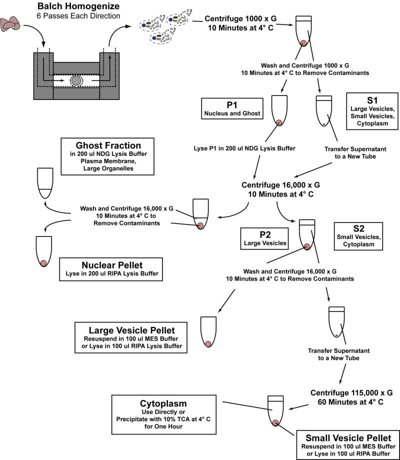
Visual overview of subcellular fractionation technique as described in the materials and methods.
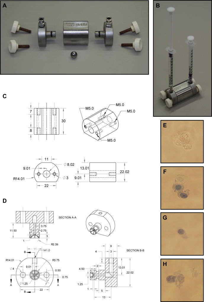
Schematic diagram of a Balch homogenizer. (A) Balch homogenizer components include a stainless steel central chamber, two stainless steel end-caps with 6.35 mm, long threaded square body female luer hubs inserted through rubber o-rings, four 25 mm M5 threaded thumb screws and a tungsten carbide ball. (B) The Balch homogenizer is constructed while submerged in water and air is evacuated prior to use. Fluid is then passed into the Balch homogenizer chamber using 1 cc syringes attached to the luer adapters. (C) Schematic diagram of the Balch homogenizer central chamber. All measurements are in millimeters. (D) Schematic diagram of the Balch homogenizer end- caps with all measurements in millimeters. Rubber O-rings must be placed into the grooves on the end-cap insert shafts to properly seal the chamber. Different luer adapters may be attached to the threaded end-caps allowing a wide variety of input sources. (E) 3T3 MEF WT cells prior to homogenization show complete exclusion of trypan blue. (F) Cells passed through an 8 μm gap show partial disruption. One cell completely excludes trypan blue while the others show varying inclusion. (G) Cells passed through a 7 μm gap show good disruption. Trypan blue readily enters the cell while the plasma membrane and cellular structure is largely intact. Further, no membrane or nuclear debris is found within the supernatant. (H) Cells passed through a 4 μm gap show signs of excessive homogenization. Nuclei are partially disrupted and separated from cell bodies while cellular debris may be seen within the supernatant.
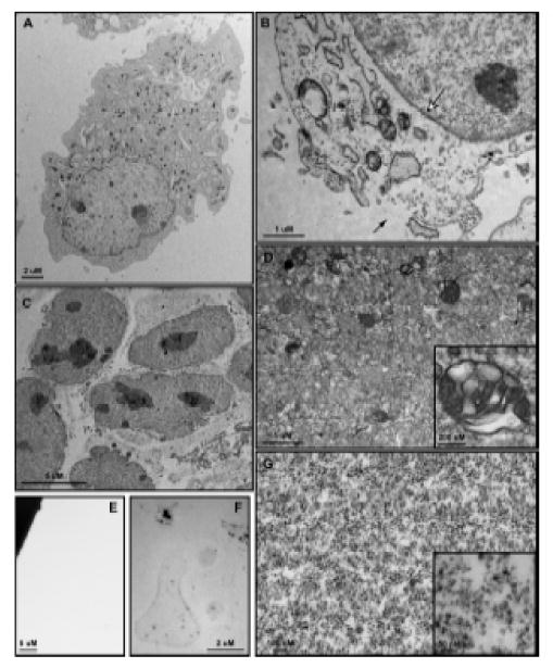
Transmission electron microscopy of 3T3 MEF WT cells and subcellular fractions. (A) Whole cells are approximately 30 – 50 μm in diameter and show a clearly defined plasma membrane with normal subcellular architecture. (B) Following Balch homogenization, small tears are formed within the plasma membrane (black arrow) through which vesicles pass during centrifugation. The nuclear envelope is entirely intact (white arrow) and subcellular structures show few signs of disturbance. (C) The nuclear fraction consists of nuclei with unbroken envelopes and attached endosplasmic reticulum, both smooth and rough. (D) The large vesicle fraction contains a heterogeneous population of membrane-bound organelles 50 nm – 0.5 μm in size. Large, multivesicular structures are well-represented within this fraction (Inset). The small vesicle fraction contains membrane-bound structures 15 nm – 25 nm in size. Electron density within this population varies widely (Inset). (F) After NDG lysis the ghost fraction contains plasma membrane micelles as well as some electron dense protein clusters. (G) No membrane or protein structures are visualized within the cytosolic fraction.
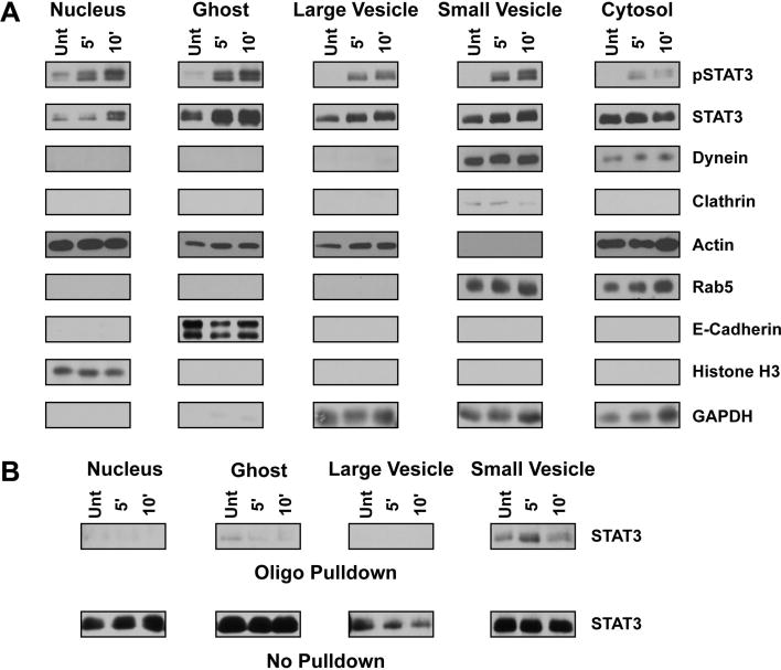
Biochemical characterization and functional assay utilizing subcellular fractions. (A) Western blot analysis of subcellular fractions isolated from 3T3 MEF WT cells treated with 10 ng/ml IL-6 for the indicated period as described within the materials and methods section. STAT3, found in all subcellular fractions, is enriched within the nuclear fraction following IL-6 treatment. Activated STAT3 may also be found within the ghost, small vesicle, and large vesicle fractions. Rab5, dynein and clathrin are all found within the small vesicle fraction, indicating this fraction is enriched with early endosomes and clathrin-coated vesicles. The complete localization of e-cadherin and histone H3 to the ghost and nuclear fractions, respectively, indicates these fractions do not contaminate the vesicular fractions. The cytosolic fraction is characterized by the presence of actin, rab5, dynein, and GAPDH, suggesting the accumulation of monomeric cytoskeletal proteins and a pool of latent proteins that interact with vesicular structures. The large vesicle fraction does not contain structures associated with early endosomes and is more thoroughly described within the text and Figure 3. (B) Western blot analysis of STAT3 DNA binding capacity following 10 ng/ml IL-6 treatment of 3T3 MEF WT cells and fraction isolation. Fractions were incubated with a biotinylated oligo containing a known STAT3 binding region and then pulled down with neutravidin agarose beads as described in the materials and methods. STAT3 isolated through oligo binding is shown in the top panel labeled ‘Oligo Pulldown’ whereas the remaining STAT3 that was unable to bind the oligo is shown in the bottom panel ‘No Pulldown’. STAT3 within the small vesicle fraction has the greatest DNA binding capacity that increases upon IL-6 treatment.
Similar articles
-
Cell Fractionation of U937 Cells in the Absence of High-speed Centrifugation.J Vis Exp. 2019 Jan 18;(143). doi: 10.3791/59022. J Vis Exp. 2019. PMID: 30735201
-
Simple and Efficient Protocol for Subcellular Fractionation of Normal and Apoptotic Cells.Cells. 2021 Apr 9;10(4):852. doi: 10.3390/cells10040852. Cells. 2021. PMID: 33918601 Free PMC article.
-
New application of a subcellular fractionation method to kidney and testis for the determination of conjugated linoleic acid in selected cell organelles of healthy and cancerous human tissues.Anal Bioanal Chem. 2005 Mar;381(6):1138-44. doi: 10.1007/s00216-004-3009-z. Epub 2005 Mar 11. Anal Bioanal Chem. 2005. PMID: 15761741
-
One-step isolation and characterization of nuclear membranes.Philos Trans R Soc Lond B Biol Sci. 1974 Jul 25;268(891):101-8. doi: 10.1098/rstb.1974.0018. Philos Trans R Soc Lond B Biol Sci. 1974. PMID: 4155085 Review. No abstract available.
-
Subcellular fractionation of adipocytes and 3T3-L1 cells.Methods Mol Biol. 2001;155:77-82. doi: 10.1385/1-59259-231-7:077. Methods Mol Biol. 2001. PMID: 11293085 Review. No abstract available.
Cited by
-
The STAT3 beacon: IL-6 recurrently activates STAT 3 from endosomal structures.Exp Cell Res. 2011 Aug 15;317(14):1955-69. doi: 10.1016/j.yexcr.2011.05.009. Epub 2011 May 18. Exp Cell Res. 2011. PMID: 21619877 Free PMC article.
-
A spatio-temporal analysis of matrix protein and nucleocapsid trafficking during vesicular stomatitis virus uncoating.PLoS Pathog. 2010 Jul 15;6(7):e1000994. doi: 10.1371/journal.ppat.1000994. PLoS Pathog. 2010. PMID: 20657818 Free PMC article.
-
Evidence for abnormal forward trafficking of AMPA receptors in frontal cortex of elderly patients with schizophrenia.Neuropsychopharmacology. 2010 Sep;35(10):2110-9. doi: 10.1038/npp.2010.87. Epub 2010 Jun 23. Neuropsychopharmacology. 2010. PMID: 20571483 Free PMC article.
References
-
- Daiss JL, Roth TF. Isolation of coated vesicles: comparative studies. Methods Enzymol. 1983;98:337–49. - PubMed
-
- Pearse BM. Isolation of coated vesicles. Methods Enzymol. 1983;98:320–6. - PubMed
-
- Morand JN, Kent C. A one-step technique for the subcellular fractionation of total cell homogenates. Anal Biochem. 1986;159:157–62. - PubMed
-
- Fuchs R, Ellinger I. Free-flow electrophoretic analysis of endosome subpopulations of rat hepatocytes. Curr Protoc Cell Biol. 2002;Chapter 3(Unit 3):11. - PubMed
Publication types
MeSH terms
Substances
Grants and funding
LinkOut - more resources
Full Text Sources
