. 2009 Oct;137(4):1367-79.e1-6.
doi: 10.1053/j.gastro.2009.07.041. Epub 2009 Jul 19.

Diet synergistically affects helicobacter pylori-induced gastric carcinogenesis in nonhuman primates

Affiliations

Diet synergistically affects helicobacter pylori-induced gastric carcinogenesis in nonhuman primates

Hui Liu et al. Gastroenterology. 2009 Oct.

Abstract

Background & aims: Gastric cancer results from a combination of Helicobacter pylori (H pylori) infection, exposure to dietary carcinogens, and predisposing genetic make-up. Because the role of these factors in gastric carcinogenesis cannot be determined readily in human beings, the present study examined the role of an oral carcinogen and H pylori infection in rhesus monkeys.

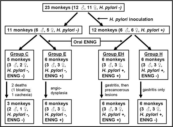
Methods: Gastroscopies were performed in 23 monkeys assigned to 4 groups: controls; nitrosating carcinogen ethyl-nitro-nitrosoguanidine administration alone; inoculation of a virulent H pylori strain alone (H); and ethyl-nitro-nitrosoguanidine in combination with H pylori (EH). Follow-up gastroscopies and biopsies were performed at 3-month intervals for 5 years for pathologic and molecular studies.

Results: Postinoculation, H and EH groups showed persistent infection and antral gastritis. Starting at 2 and 5 years, respectively, gastric intestinal metaplasia and intraepithelial neoplasia developed in 3 EH monkeys but in no other groups. Transcriptional analysis of biopsy specimens at 5 years revealed group-specific expression profiles, with striking changes in EH monkeys, plus a neoplasia-specific expression profile characterized by changes in multiple cancer-associated genes. Importantly, this neoplastic profile was evident in nonneoplastic mucosa, suggesting that the identified genes may represent markers preceding cancer.

Conclusions: Gastric intraglandular neoplasia is induced in primates when H pylori infection is associated with consumption of a carcinogen similar to the nitrosamines found in pickled vegetables, suggesting that H pylori and the carcinogen synergistically induce gastric neoplasia in primates.


Figures

Figure 1
Figure 1
Experimental design, time course and outcome of the study.
Figure 2
Figure 2
Effect of H. pylori inoculation on H. pylori load (copies/100 ng total RNA) and gastritis scores in the gastric antrum (Mean±SEM). (a) “Preinoc” indicates the uninfected status of the animals at the initiation of the study and is given as a reference. Infected animals were dosed with 108 H. pylori CFU/ml and colonization burden was monitored over time. (b) Gastritis scores were calculated using the updated Sydney System. Note that gastritis scores were similar in the H and EH groups although infectious load was lower in the EH group.
Figure 3
Figure 3
Illustration of endoscopic views of the stomach of (a) Control monkey; (b) H. pylori infected monkey with gastroscopic nodularity; and (c) EH animal with a polyp (white arrow) and telangectasias (black arrow).
Figure 4
Figure 4
Histopathology of gastric mucosa in the four groups of monkeys illustrating (a) normal antrum in control monkey at 5-year; (b) antrum from E monkey at 5-year [grade 0 gastritis; hyperchromatic epithelial cells with bigger, more spherical nuclei than in controls, and enlarged lamina propria with pink matrix]; (c) grade 3 gastritis and grade 2 focal atrophy of the antrum in H monkey at 4-year [note intraepithelial lymphocytes]; (d) grade 3 gastritis and grade 1 focal atrophy of the corpus in EH monkey at 3-year; (e) grade 3 gastritis and grade 2 focal atrophy of the corpus in an EH monkey at 3-year [note dilated capillaries in lamina propria]; (f) grade 3 gastritis and grade 1 focal atrophy of the antrum in an EH monkey at 5-year; (g) complete gastric intestinal metaplasia by H&E stain in an EH monkey at 3-year [G: Goblet cells; thin arrows: Paneth cells; arrowheads: absorptive intestinal cells] and (h) by Genta stain [Alcian blue staining of goblet cells]; insert: CDX2-stained nuclei of goblet cells (i) histology of polyp resected at 5-year [low magnification of polyp showing abnormal histology in area delimited by an imaginary line joining the two arrowheads]; and (j) high magnification of region delimited by the box in (i) demonstrating intraepithelial neoplasia with presence of hyperchromatic stratified nuclei (arrowheads) and mitosis (thin arrows).
Figure 5
Figure 5
Effect of antibiotics and H. pylori on host gene expression. (a-c) “Arrival” denotes initial naturally infected status, i.e. before the monkeys were treated with antibiotics. “Preinoc” indicates the uninfected status of the animals at the initiation of the study and is given as a reference. mRNA expression of IL-1β, IL-8, and MGMT as determined by real-time RT-PCR in C, E, H and EH monkeys (Means ± SEM). Note that, similar to the dynamics of gastritis illustrated in Fig. 2, expression of the three genes decreases after antibiotic treatment and increases at one-year after H. pylori inoculation in EH and H monkeys. However, at later time points, expression of the inflammatory cytokines remains elevated whereas expression of the DNA repair gene MGMT returns to levels similar to those before H. pylori inoculation. In the C and E groups there was no significant difference for IL-1β, IL8, or MGMT expression as compared to the initial levels. (d) Cluster Diagram of the genes showing statistically significant differences in gastric biopsies harvested at 5 years. Letters indicate the relative treatment groups and numbers indicate the particular animal within the indicated group. Red and green colors represent increase or decreased expression, respectively, relative to the reference sample. Note the dendogram shows two major nodes that segregate the majority of C and H samples from E and EH samples. (e) Dendogram showing clustering pattern of C and EH arrays as supervised by the 83-gene neoplastic signature in gastric biopsies harvested at 5 years. Note that all neoplastic samples segregate from non-neoplastic EH and Control samples.

Similar articles

Cited by

References

    1. Crew KD, Neugut AI. Epidemiology of gastric cancer. World J Gastroenterol. 2006;12:354–362. - PMC - PubMed
    1. Parkin DM, Bray F, Ferlay J, Pisani P. Global cancer statistics, 2002. CA Cancer J Clin. 2005;55:74–108. - PubMed
    1. Haenszel W, Correa P. Developments in the epidemiology of stomach cancer over the past decade. Cancer Res. 1975;35:3452–3459. - PubMed
    1. Marshall BJ, Warren JR. Unidentified curved bacilli in the stomach of patients with gastritis and peptic ulceration. Lancet. 1984;1:1311–1315. - PubMed
    1. Parsonnet J, Friedman GD, Vandersteen DP, Chang Y, Vogelman JH, Orentreich N, Sibley RK. Helicobacter pylori infection and the risk of gastric carcinoma. N Engl J Med. 1991;325:1127–1131. - PubMed

MeSH terms