

HER family receptor abnormalities in lung cancer brain metastases and corresponding primary tumors
- PMID: 19622585
- PMCID: PMC3372920
- DOI: 10.1158/1078-0432.CCR-08-2921
HER family receptor abnormalities in lung cancer brain metastases and corresponding primary tumors
Abstract
Purpose: To compare the characteristics of deregulation of HER receptors and their ligands between primary tumor and corresponding brain metastases of non-small cell lung carcinoma (NSCLC).
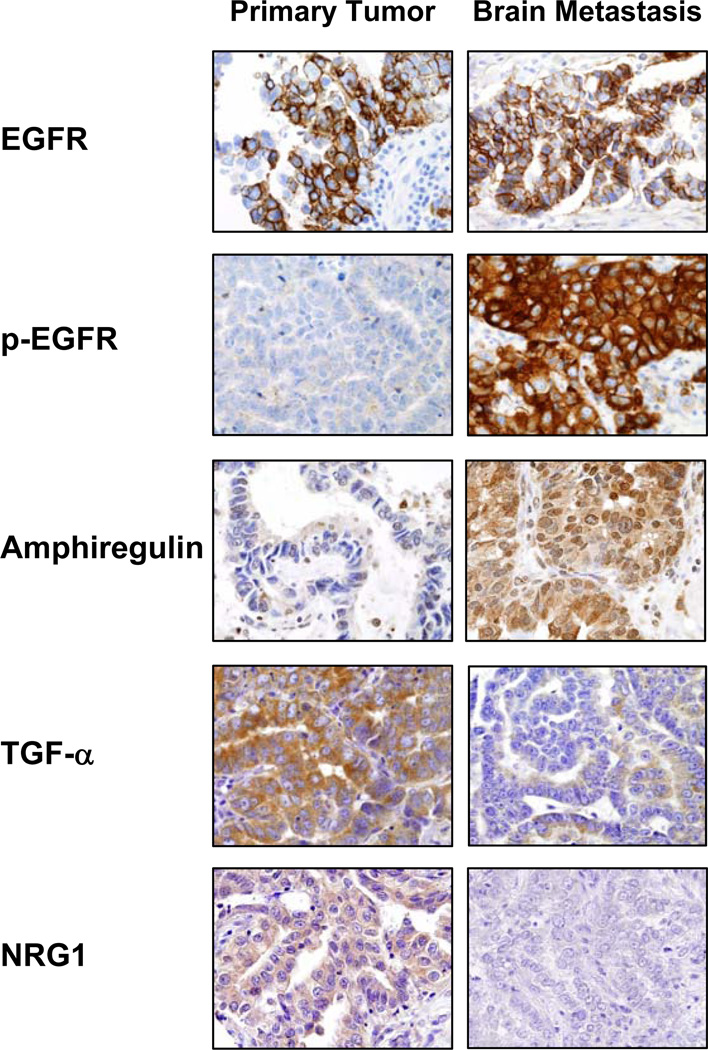
Experimental design: Fifty-five NSCLC primary tumors and corresponding brain metastases specimens were examined for the immunohistochemical expression of epidermal growth factor receptor (EGFR), phosphorylated EGFR, Her2, Her3, and phosphorylated Her3, and their ligands EGF, transforming growth factor-alpha, amphiregulin, epiregulin, betacellulin, heparin-binding EGFR-like growth factor, neuregulin (NRG) 1, and NRG2. Analysis of EGFR copy number using fluorescence in situ hybridization and mutation by PCR-based sequencing was also done.
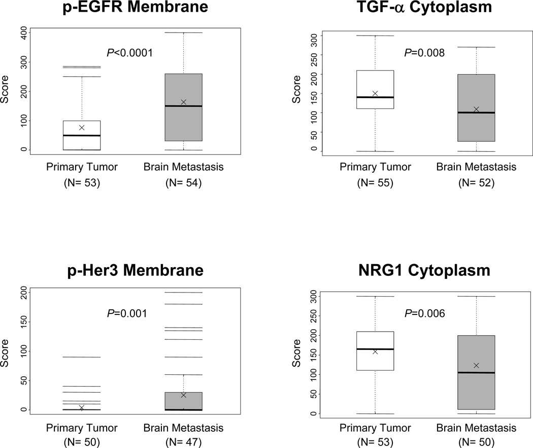
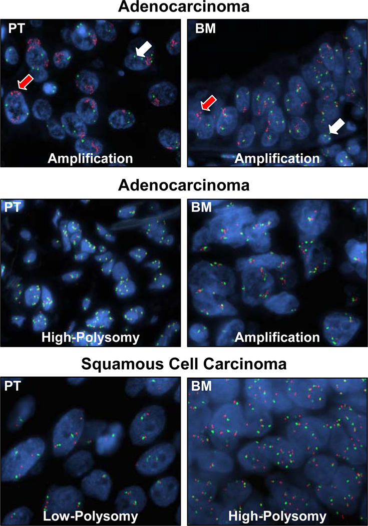
Results: Metastases showed significantly higher immunohistochemical expression of EGF (membrane: brain metastases 66.0 versus primary tumors 48.5; P = 0.027; nucleus: brain metastases 92.2 versus 67.4; P = 0.008), amphiregulin (nucleus: brain metastases 53.7 versus primary tumors 33.7; P = 0.019), phosphorylated EGFR (membrane: brain metastases 161.5 versus primary tumors 76.0; P < 0.0001; cytoplasm: brain metastases 101.5 versus primary tumors 55.9; P = 0.014), and phosphorylated Her3 (membrane: brain metastases 25.0 versus primary tumors 3.7; P = 0.001) than primary tumors did. Primary tumors showed significantly higher expression of cytoplasmic transforming growth factor-alpha(primary tumors 149.8 versus brain metastases 111.3; P = 0.008) and NRG1 (primary tumors 158.5 versus brain metastases 122.8; P = 0.006). In adenocarcinomas, a similar high frequency of EGFR copy number gain (high polysomy and amplification) was detected in primary (65%) and brain metastasis (63%) sites. However, adenocarcinoma metastases (30%) showed higher frequency of EGFR amplification than corresponding primary tumors (10%). Patients whose primary tumors showed EGFR amplification tended to develop brain metastases at an earlier time point.
Conclusions: Our findings suggest that NSCLC brain metastases have some significant differences in HER family receptor-related abnormalities from primary lung tumors.
Figures
References
-
- Travis WD, Brambilla E, Muller-Hermelink HK, Harris CC. Tumours of the lung. In: Travis WD, Brambilla E, Muller-Hermelink HK, Harris CC, editors. Pathology and Genetics: Tumours of the Lung, Pleura, Thymus and Heart. Lyon: International Agency for Research on Cancer (IARC); 2004. pp. 9–124.
-
- Stuschke M, Eberhardt W, Pottgen C, et al. Prophylactic cranial irradiation in locally advanced non-small-cell lung cancer after multimodality treatment: long-term follow-up and investigations of late neuropsychologic effects. J Clin Oncol. 1999;17:2700–2709. - PubMed
-
- Omuro AM, Kris MG, Miller VA, et al. High incidence of disease recurrence in the brain and leptomeninges in patients with nonsmall cell lung carcinoma after response to gefitinib. Cancer. 2005;103:2344–2348. - PubMed
-
- Mamon HJ, Yeap BY, Janne PA, et al. High risk of brain metastases in surgically staged IIIA non-small-cell lung cancer patients treated with surgery, chemotherapy, and radiation. J Clin Oncol. 2005;23:1530–1537. - PubMed
-
- Chen AM, Jahan TM, Jablons DM, Garcia J, Larson DA. Risk of cerebral metastases and neurological death after pathological complete response to neoadjuvant therapy for locally advanced nonsmall-cell lung cancer: clinical implications for the subsequent management of the brain. Cancer. 2007;109:1668–1675. - PubMed
Publication types
MeSH terms
Substances
Grants and funding
LinkOut - more resources
Full Text Sources
Other Literature Sources
Medical
Research Materials
Miscellaneous
