

TMS activation of interhemispheric pathways between the posterior parietal cortex and the contralateral motor cortex
- PMID: 19622612
- PMCID: PMC2754365
- DOI: 10.1113/jphysiol.2009.174086
TMS activation of interhemispheric pathways between the posterior parietal cortex and the contralateral motor cortex
Abstract
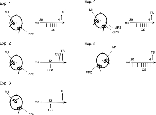
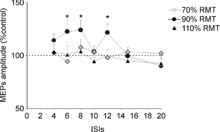
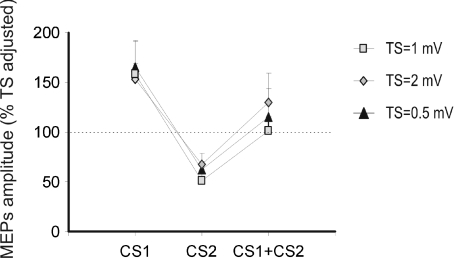
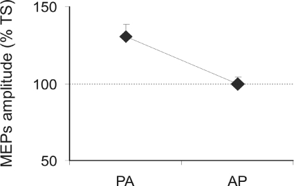
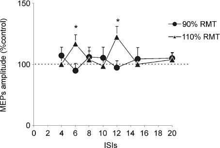
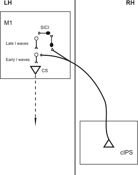
Using a twin coil transcranial magnetic stimulation (tc-TMS) approach we have previously demonstrated that facilitation may be detected in the primary motor cortex (M1) following stimulation over the ipsilateral caudal intraparietal sulcus (cIPS). Here we tested the interhemispheric interactions between the IPS and the contralateral motor cortex (M1). We found that conditioning the right cIPS facilitated contralateral M1 when the conditioning stimulus had an intensity of 90% resting motor threshold (RMT) but not at 70% or 110% RMT. Facilitation was maximal when the interstimulus interval (ISI) between cIPS and M1 was 6 or 12 ms. These facilitatory effects were mediated by interactions with specific groups of interneurons in the contralateral M1. In fact, short intracortical inhibition (SICI) was reduced following cIPS stimulation. Moreover, additional comparison of facilitation of responses evoked by anterior-posterior versus posterior-anterior stimulation of M1 suggested that facilitation was more effective on early I1/I2 circuits than on I3 circuits. In contrast to these effects, stimulation of anterior IPS (aIPS) at 90% RMT induced inhibition, instead of facilitation, of contralateral M1 at ISIs of 10-12 ms. Finally, we found similar facilitation between left cIPS and right M1 although the conditioning stimuli had to have a higher intensity compared with stimulation of right cIPS (110% instead of 90% RMT). These findings demonstrate that different subregions of the posterior parietal cortex (PPC) in humans exert both facilitatory and inhibitory effects towards the contralateral primary motor cortex. These corticocortical projections could contribute to a variety of motor tasks such as bilateral manual coordination, movement planning in space and grasping.
Figures
Similar articles
-
Focal stimulation of the posterior parietal cortex increases the excitability of the ipsilateral motor cortex.J Neurosci. 2007 Jun 20;27(25):6815-22. doi: 10.1523/JNEUROSCI.0598-07.2007. J Neurosci. 2007. PMID: 17581969 Free PMC article.
-
Magnetic stimulation of human premotor or motor cortex produces interhemispheric facilitation through distinct pathways.J Physiol. 2006 May 1;572(Pt 3):857-68. doi: 10.1113/jphysiol.2006.104901. J Physiol. 2006. PMID: 16497712 Free PMC article. Clinical Trial.
-
Interhemispheric interactions between the right angular gyrus and the left motor cortex: a transcranial magnetic stimulation study.J Neurophysiol. 2021 Apr 1;125(4):1236-1250. doi: 10.1152/jn.00642.2020. Epub 2021 Feb 24. J Neurophysiol. 2021. PMID: 33625938
-
TMS investigations into the task-dependent functional interplay between human posterior parietal and motor cortex.Behav Brain Res. 2009 Sep 14;202(2):147-52. doi: 10.1016/j.bbr.2009.03.023. Epub 2009 Mar 28. Behav Brain Res. 2009. PMID: 19463695 Review.
-
Beyond grasping: representation of action in human anterior intraparietal sulcus.Neuroimage. 2007;36 Suppl 2(Suppl 2):T77-86. doi: 10.1016/j.neuroimage.2007.03.026. Epub 2007 Mar 28. Neuroimage. 2007. PMID: 17499173 Free PMC article. Review.
Cited by
-
Dissociating Arithmetic Operations in the Parietal Cortex Using 1 Hz Repetitive Transcranial Magnetic Stimulation: The Importance of Strategy Use.Front Hum Neurosci. 2020 Jul 16;14:271. doi: 10.3389/fnhum.2020.00271. eCollection 2020. Front Hum Neurosci. 2020. PMID: 32765240 Free PMC article.
-
1 Hz rTMS of the left posterior parietal cortex (PPC) modifies sensorimotor timing.Neuropsychologia. 2012 Dec;50(14):3729-35. doi: 10.1016/j.neuropsychologia.2012.10.020. Epub 2012 Oct 26. Neuropsychologia. 2012. PMID: 23103789 Free PMC article.
-
Paired associative stimulation applied to the cortex can increase resting-state functional connectivity: A proof of principle study.Neurosci Lett. 2022 Jul 27;784:136753. doi: 10.1016/j.neulet.2022.136753. Epub 2022 Jun 23. Neurosci Lett. 2022. PMID: 35753613 Free PMC article.
-
Role of the Contralesional Hemisphere in Post-Stroke Recovery of Upper Extremity Motor Function.Front Neurol. 2015 Oct 16;6:214. doi: 10.3389/fneur.2015.00214. eCollection 2015. Front Neurol. 2015. PMID: 26528236 Free PMC article. Review.
-
The illusion of changed position and movement from vibrating one arm is altered by vision or movement of the other arm.J Physiol. 2010 Aug 1;588(Pt 15):2789-800. doi: 10.1113/jphysiol.2010.192336. Epub 2010 Jun 14. J Physiol. 2010. PMID: 20547672 Free PMC article.
References
-
- Amassian VE, Stewart M. Motor cortical and other cortical interneuronal networks that generate very high frequency waves. Suppl Clin Neurophysiol. 2003;56:119–142. - PubMed
-
- Andersen RA, Buneo CA. Intentional maps in posterior parietal cortex. Annu Rev Neurosci. 2002;25:189–220. - PubMed
-
- Binkofski F, Buccino G, Posse S, Seitz RJ, Rizzolatti G, Freund H. A frontoparietal circuit for object manipulation in man: evidence from an fMRI-study. Eur J Neurosci. 1999;11:3276–3286. - PubMed
-
- Caminiti R, Ferraina S, Johnson PB. The sources of visual information to the primate frontal lobe: a novel role for the superior parietal lobule. Cereb Cortex. 1996;6:319–328. - PubMed
Publication types
MeSH terms
Grants and funding
LinkOut - more resources
Full Text Sources