. 2009 Aug 11;106(32):13307-133310.
doi: 10.1073/pnas.0902312106. Epub 2009 Jul 21.

The titin-telethonin complex is a directed, superstable molecular bond in the muscle Z-disk

Affiliations

The titin-telethonin complex is a directed, superstable molecular bond in the muscle Z-disk

Morten Bertz et al. Proc Natl Acad Sci U S A. .

Abstract

Mechanical stability of bonds and protein interactions has recently become accessible through single molecule mechanical experiments. So far, mechanical information about molecular bond mechanics has been largely limited to a single direction of force application. However, mechanical force acts as a vector in space and hence mechanical stability should depend on the direction of force application. In skeletal muscle, the giant protein titin is anchored in the Z-disk by telethonin. Much of the structural integrity of the Z-disk hinges upon the titin-telethonin bond. In this paper we show that the complex between the muscle proteins titin and telethonin forms a highly directed molecular bond. It is designed to resist ultra-high forces if they are applied in the direction along which it is loaded under physiological conditions, while it breaks easily along other directions. Highly directed molecular bonds match in an ideal way the requirements of tissues subject to mechanical stress.


Conflict of interest statement

The authors declare no conflict of interest.

Figures

Fig. 1.
Fig. 1.
(A) Structure of the antiparallel titin-telethonin complex (pdb: 1YA5) (10). The first 2 N-terminal domains [Z1 (dark) and Z2 (light)] of 2 titin molecules [chain A (green) and chain B (blue)] are assembled into a palindromic complex by telethonin (pink). (B) Schematic overview of the sarcomere and its 3 major filaments: actin, myosin, and titin. Only one-half of the sarcomere is shown. The titin-telethonin complex located in the Z-disk is shown as a space-filling model. (C) Typical force-extension traces of the titin-telethonin complex pulled at its C termini (Z1Z2TC-C): The structural model illustrates the pulling geometry and the location of the cysteine crosslinks (yellow bars, compare Fig. S1) necessary to obtain a clear fingerprint of the rupture of the complex. Unfolding events of the ubiquitin handles (gray filled circles in the structural model) are colored gray in the unfolding traces. WLC curves fit to the rupture of the complex (colored part of the traces) are shown as dashed lines with contour length increases indicated above the curves. (D) Force histogram of the initial rupture event of the complex (green circle in C).
Fig. 2.
Fig. 2.
(A) Force-extension traces of the Z1Z2 fragment from titin flanked by ubiquitin domains (gray filled circles). Unfolding events of the ubiqutin handles are colored in gray in the traces. Unfolding events of the titin fragment are colored in green. Dashed lines indicate WLC fits with the contour length increases shown above the curves. (B) Force histogram of the mechanical unfolding of the Z1Z2 domain pair. (C) Force-extension traces of the inverted pulling geometry of the titin-telethonin complex (Z1Z2TN-N): The structural model illustrates the architecture of the construct. Cysteine crosslinks are shown as yellow bars (compare Fig. S1), ubiquitin handles as gray filled circles. The rupture of Z1Z2TN-N is indicated by the various colors in the traces. WLC fits to the data and contour length increases are show in the left panel. (D) Force histogram of the initial rupture event of Z1Z2TN-N.
Fig. 3.
Fig. 3.
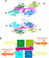
(A) Top: Structure of the titin-telethonin complex with part of the hydrogen bond network (black lines) stabilizing the distal Z2 domains when load is applied at the C termini. Bottom: Back view of the titin-telethonin complex. The N termini of the proximal Z1 domains do not form a tight hydrogen bond network with telethonin. (B) The titin-telethonin complex is a directed, highly stable molecular bond. Load applied to the C termini, as is the case in the muscle during passive stretching, requires high forces to dissociate the complex. If loaded at the N termini, the complex unravels readily.

Comment in

  • A new direction for titin pulling.
    Rock RS. Rock RS. Proc Natl Acad Sci U S A. 2009 Aug 11;106(32):13149-50. doi: 10.1073/pnas.0906989106. Epub 2009 Aug 5. Proc Natl Acad Sci U S A. 2009. PMID: 19666561 Free PMC article. No abstract available.

Similar articles

Cited by

References

    1. Tskhovrebova L, Trinick J. Titin: Properties and family relationships. Nat Rev Mol Cell Biol. 2003;4:679–689. - PubMed
    1. Labeit S, Kolmerer B. Titins: Giant proteins in charge of muscle ultrastructure and elasticity. Science. 1995;270:293–296. - PubMed
    1. Maruyama K. Connectin/titin, giant elastic protein of muscle. FASEB J. 1997;11:341–345. - PubMed
    1. Li H, et al. Reverse engineering of the giant muscle protein titin. Nature. 2002;418:998–1002. - PubMed
    1. Mayans O, et al. Structural basis for activation of the titin kinase domain during myofibrillogenesis. Nature. 1998;395:863–869. - PubMed

Publication types

LinkOut - more resources