

A Ca2(+ )release-activated Ca2(+) (CRAC) modulatory domain (CMD) within STIM1 mediates fast Ca2(+)-dependent inactivation of ORAI1 channels
- PMID: 19622747
- PMCID: PMC2757196
- DOI: 10.1074/jbc.C109.024083
A Ca2(+ )release-activated Ca2(+) (CRAC) modulatory domain (CMD) within STIM1 mediates fast Ca2(+)-dependent inactivation of ORAI1 channels
Abstract
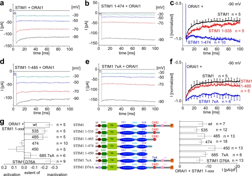
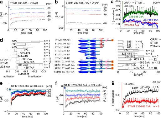
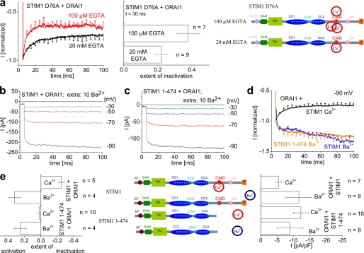
STIM1 and ORAI1, the two limiting components in the Ca(2+) release-activated Ca(2+) (CRAC) signaling cascade, have been reported to interact upon store depletion, culminating in CRAC current activation. We have recently identified a modulatory domain between amino acids 474 and 485 in the cytosolic part of STIM1 that comprises 7 negatively charged residues. A STIM1 C-terminal fragment lacking this domain exhibits enhanced interaction with ORAI1 and 2-3-fold higher ORAI1/CRAC current densities. Here we focused on the role of this CRAC modulatory domain (CMD) in the fast inactivation of ORAI1/CRAC channels, utilizing the whole-cell patch clamp technique. STIM1 mutants either with C-terminal deletions including CMD or with 7 alanines replacing the negative amino acids within CMD gave rise to ORAI1 currents that displayed significantly reduced or even abolished inactivation when compared with STIM1 mutants with preserved CMD. Consistent results were obtained with cytosolic C-terminal fragments of STIM1, both in ORAI1-expressing HEK 293 cells and in RBL-2H3 mast cells containing endogenous CRAC channels. Inactivation of the latter, however, was much more pronounced than that of ORAI1. The extent of inactivation of ORAI3 channels, which is also considerably more prominent than that of ORAI1, was also substantially reduced by co-expression of STIM1 constructs missing CMD. Regarding the dependence of inactivation on Ca(2+), a decrease in intracellular Ca(2+) chelator concentrations promoted ORAI1 current fast inactivation, whereas Ba(2+) substitution for extracellular Ca(2+) completely abrogated it. In summary, CMD within the STIM1 cytosolic part provides a negative feedback signal to Ca(2+) entry by triggering fast Ca(2+)-dependent inactivation of ORAI/CRAC channels.
Figures
Similar articles
-
A Cytosolic Homomerization and a Modulatory Domain within STIM1 C Terminus Determine Coupling to ORAI1 Channels.J Biol Chem. 2009 Mar 27;284(13):8421-6. doi: 10.1074/jbc.C800229200. Epub 2009 Feb 3. J Biol Chem. 2009. PMID: 19189966 Free PMC article.
-
Store-Independent Orai Channels Regulated by STIM.In: Kozak JA, Putney JW Jr, editors. Calcium Entry Channels in Non-Excitable Cells. Boca Raton (FL): CRC Press/Taylor & Francis; 2018. Chapter 11. In: Kozak JA, Putney JW Jr, editors. Calcium Entry Channels in Non-Excitable Cells. Boca Raton (FL): CRC Press/Taylor & Francis; 2018. Chapter 11. PMID: 30299650 Free Books & Documents. Review.
-
STIM1 and calmodulin interact with Orai1 to induce Ca2+-dependent inactivation of CRAC channels.Proc Natl Acad Sci U S A. 2009 Sep 8;106(36):15495-500. doi: 10.1073/pnas.0906781106. Epub 2009 Aug 21. Proc Natl Acad Sci U S A. 2009. PMID: 19706428 Free PMC article.
-
Store-dependent and -independent modes regulating Ca2+ release-activated Ca2+ channel activity of human Orai1 and Orai3.J Biol Chem. 2008 Jun 20;283(25):17662-71. doi: 10.1074/jbc.M801536200. Epub 2008 Apr 17. J Biol Chem. 2008. PMID: 18420579 Free PMC article.
-
The STIM/Orai coupling machinery.Channels (Austin). 2008 Jul-Aug;2(4):261-8. doi: 10.4161/chan.2.4.6705. Epub 2008 Jul 21. Channels (Austin). 2008. PMID: 18769136 Review.
Cited by
-
STIM proteins: dynamic calcium signal transducers.Nat Rev Mol Cell Biol. 2012 Sep;13(9):549-65. doi: 10.1038/nrm3414. Nat Rev Mol Cell Biol. 2012. PMID: 22914293 Free PMC article. Review.
-
Store-Operated Calcium Channels: From Function to Structure and Back Again.Cold Spring Harb Perspect Biol. 2020 May 1;12(5):a035055. doi: 10.1101/cshperspect.a035055. Cold Spring Harb Perspect Biol. 2020. PMID: 31570335 Free PMC article. Review.
-
Molecular physiology and pathophysiology of stromal interaction molecules.Exp Biol Med (Maywood). 2018 Mar;243(5):451-472. doi: 10.1177/1535370218754524. Epub 2018 Jan 24. Exp Biol Med (Maywood). 2018. PMID: 29363328 Free PMC article. Review.
-
Orai1 and STIM1 in ER/PM junctions: roles in pancreatic cell function and dysfunction.Am J Physiol Cell Physiol. 2016 Mar 15;310(6):C414-22. doi: 10.1152/ajpcell.00349.2015. Epub 2016 Jan 6. Am J Physiol Cell Physiol. 2016. PMID: 26739495 Free PMC article. Review.
-
The intracellular loop of Orai1 plays a central role in fast inactivation of Ca2+ release-activated Ca2+ channels.J Biol Chem. 2010 Feb 12;285(7):5066-75. doi: 10.1074/jbc.M109.072736. Epub 2009 Dec 10. J Biol Chem. 2010. PMID: 20007711 Free PMC article.
References
Publication types
MeSH terms
Substances
LinkOut - more resources
Full Text Sources
Miscellaneous
