

Tumor vascular changes mediated by inhibition of oncogenic signaling
- PMID: 19622766
- PMCID: PMC2825046
- DOI: 10.1158/0008-5472.CAN-09-0657
Tumor vascular changes mediated by inhibition of oncogenic signaling
Abstract
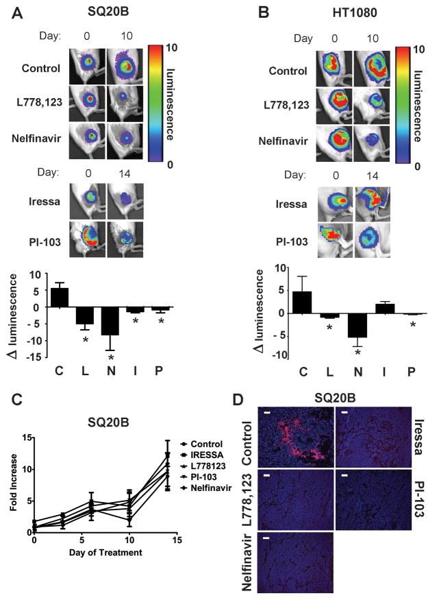
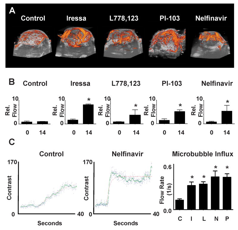
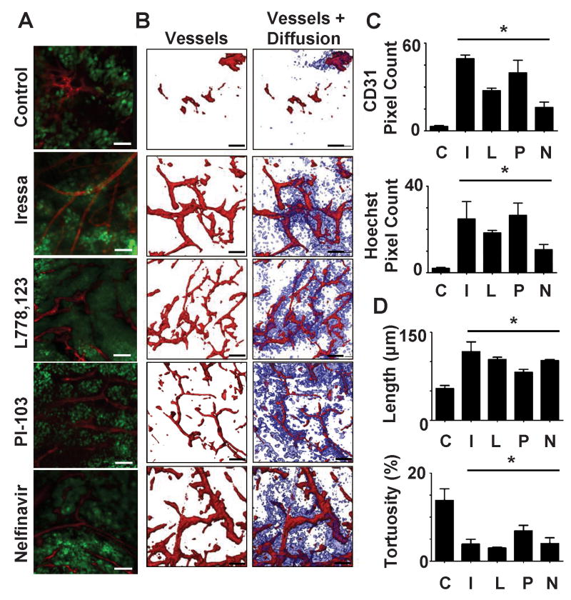
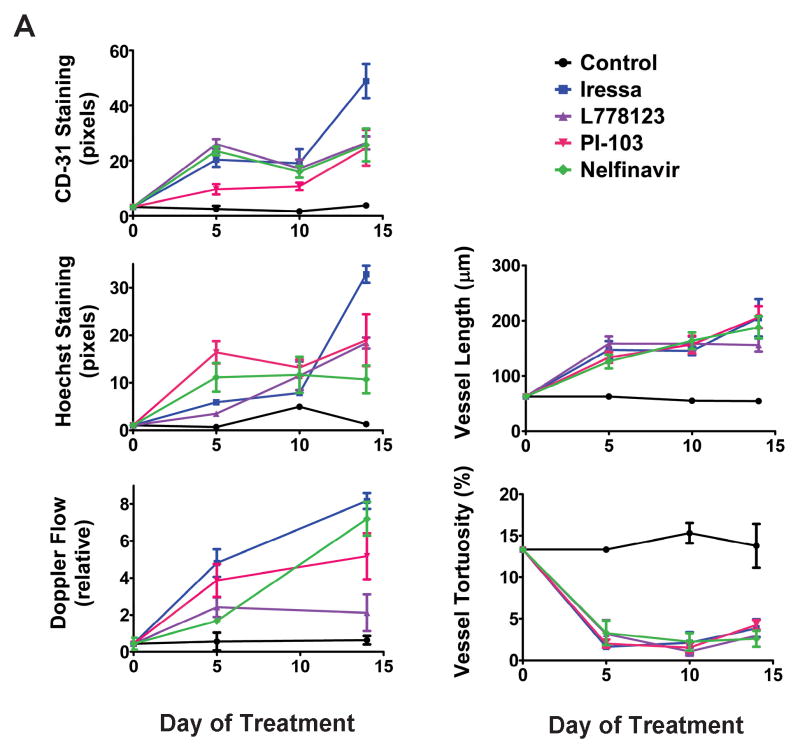
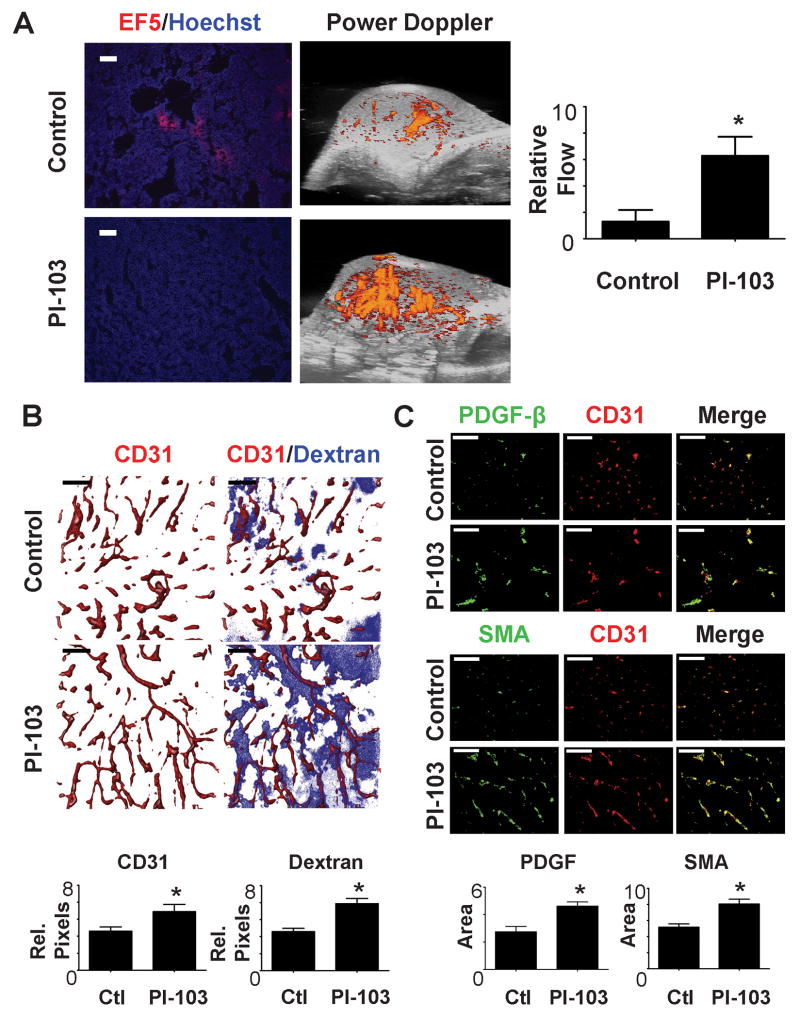
Many inhibitors of the epidermal growth factor receptor (EGFR)-RAS-phosphatidylinositol 3-kinase (PI3K)-AKT signaling pathway are in clinical use or under development for cancer therapy. Here, we show that treatment of mice bearing human tumor xenografts with inhibitors that block EGFR, RAS, PI3K, or AKT resulted in prolonged and durable enhancement of tumor vascular flow, perfusion, and decreased tumor hypoxia. The vessels in the treated tumors had decreased tortuosity and increased internodal length accounting for the functional alterations. Inhibition of tumor growth cannot account for these results, as the drugs were given at doses that did not alter tumor growth. The tumor cell itself was an essential target, as HT1080 tumors that lack EGFR did not respond to an EGFR inhibitor but did respond with vascular alterations to RAS or PI3K inhibition. We extended these observations to spontaneously arising tumors in MMTV-neu mice. These tumors also responded to PI3K inhibition with decreased tumor hypoxia, increased vascular flow, and morphologic alterations of their vessels, including increased vascular maturity and acquisition of pericyte markers. These changes are similar to the vascular normalization that has been described after the antiangiogenic treatment of xenografts. One difficulty in the use of vascular normalization as a therapeutic strategy has been its limited duration. In contrast, blocking tumor cell RAS-PI3K-AKT signaling led to persistent vascular changes that might be incorporated into clinical strategies based on improvement of vascular flow or decreased hypoxia. These results indicate that vascular alterations must be considered as a consequence of signaling inhibition in cancer therapy.
Figures
Similar articles
-
TANKYRASE Inhibition Enhances the Antiproliferative Effect of PI3K and EGFR Inhibition, Mutually Affecting β-CATENIN and AKT Signaling in Colorectal Cancer.Mol Cancer Res. 2018 Mar;16(3):543-553. doi: 10.1158/1541-7786.MCR-17-0362. Epub 2017 Dec 8. Mol Cancer Res. 2018. PMID: 29222171
-
Radiosensitization of epidermal growth factor receptor/HER2-positive pancreatic cancer is mediated by inhibition of Akt independent of ras mutational status.Clin Cancer Res. 2010 Feb 1;16(3):912-23. doi: 10.1158/1078-0432.CCR-09-1324. Epub 2010 Jan 26. Clin Cancer Res. 2010. PMID: 20103665 Free PMC article.
-
Cotargeting of epidermal growth factor receptor and PI3K overcomes PI3K-Akt oncogenic dependence in pancreatic ductal adenocarcinoma.Clin Cancer Res. 2014 Aug 1;20(15):4047-58. doi: 10.1158/1078-0432.CCR-13-3377. Epub 2014 Jun 3. Clin Cancer Res. 2014. PMID: 24895459
-
Inhibiting the RAS-PI3K pathway in cancer therapy.Enzymes. 2013;34 Pt. B:107-36. doi: 10.1016/B978-0-12-420146-0.00005-6. Epub 2013 Nov 7. Enzymes. 2013. PMID: 25034102 Review.
-
Signaling inhibition with radiation in colorectal cancer: clinical trials.Semin Oncol. 2003 Jun;30(3 Suppl 6):56-67. doi: 10.1016/s0093-7754(03)00126-x. Semin Oncol. 2003. PMID: 12802796 Review.
Cited by
-
Vascular normalisation as the stepping stone into tumour microenvironment transformation.Br J Cancer. 2021 Aug;125(3):324-336. doi: 10.1038/s41416-021-01330-z. Epub 2021 Apr 7. Br J Cancer. 2021. PMID: 33828258 Free PMC article. Review.
-
Hypoxia-inducing factors as master regulators of stemness properties and altered metabolism of cancer- and metastasis-initiating cells.J Cell Mol Med. 2013 Jan;17(1):30-54. doi: 10.1111/jcmm.12004. Epub 2013 Jan 10. J Cell Mol Med. 2013. PMID: 23301832 Free PMC article.
-
Targeting radiation-resistant hypoxic tumour cells through ATR inhibition.Br J Cancer. 2012 Jul 10;107(2):291-9. doi: 10.1038/bjc.2012.265. Epub 2012 Jun 19. Br J Cancer. 2012. PMID: 22713662 Free PMC article.
-
Modulating the tumor microenvironment to increase radiation responsiveness.Cancer Biol Ther. 2009 Nov;8(21):1994-2001. doi: 10.4161/cbt.8.21.9988. Epub 2009 Nov 3. Cancer Biol Ther. 2009. PMID: 19823031 Free PMC article. Review.
-
Dasatinib promotes the expansion of a therapeutically superior T-cell repertoire in response to dendritic cell vaccination against melanoma.Oncoimmunology. 2014 Jan 1;3(1):e27589. doi: 10.4161/onci.27589. Epub 2014 Feb 27. Oncoimmunology. 2014. PMID: 24734217 Free PMC article.
References
-
- Boucher Y, Baxter LT, Jain RK. Interstitial pressure gradients in tissue-isolated and subcutaneous tumors: implications for therapy. Cancer Research. 1990;50(15):4478–84. - PubMed
-
- Vaupel P. Tumor microenvironmental physiology and its implications for radiation oncology. Semin Radiat Oncol. 2004;14(3):198–206. - PubMed
-
- Cosse JP, Michiels C. Tumour hypoxia affects the responsiveness of cancer cells to chemotherapy and promotes cancer progression. Current Medicinal Chemistry - Anti-Cancer Agents. 2008;8(7):790–7. - PubMed
Publication types
MeSH terms
Substances
Grants and funding
LinkOut - more resources
Full Text Sources
Molecular Biology Databases
Research Materials
Miscellaneous
