

In vivo kinematics of the tibiotalar joint after lateral ankle instability
- PMID: 19622791
- PMCID: PMC2891039
- DOI: 10.1177/0363546509337578
In vivo kinematics of the tibiotalar joint after lateral ankle instability
Abstract
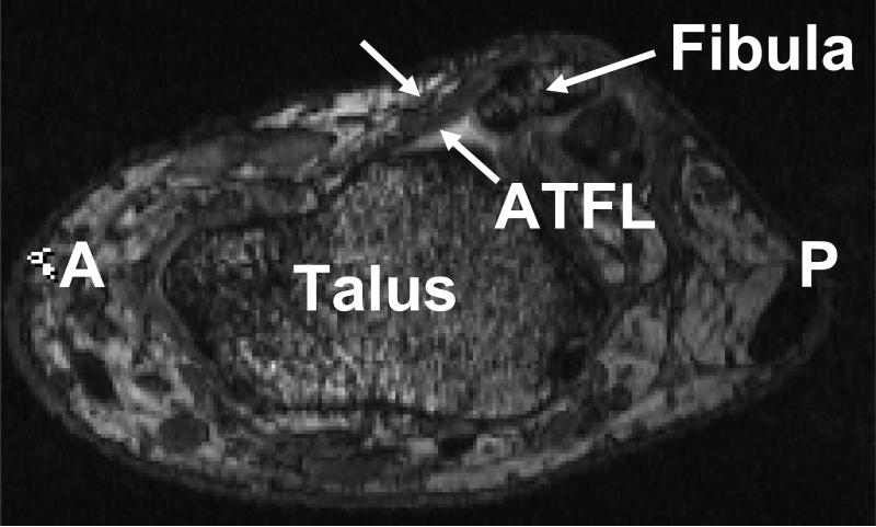
Background: Previous studies have suggested that injury to the anterior talofibular ligament (ATFL) may be linked to altered kinematics and the development of osteoarthritis of the ankle joint. However, the effects of ATFL injury on the in vivo kinematics of the ankle joint are unclear.
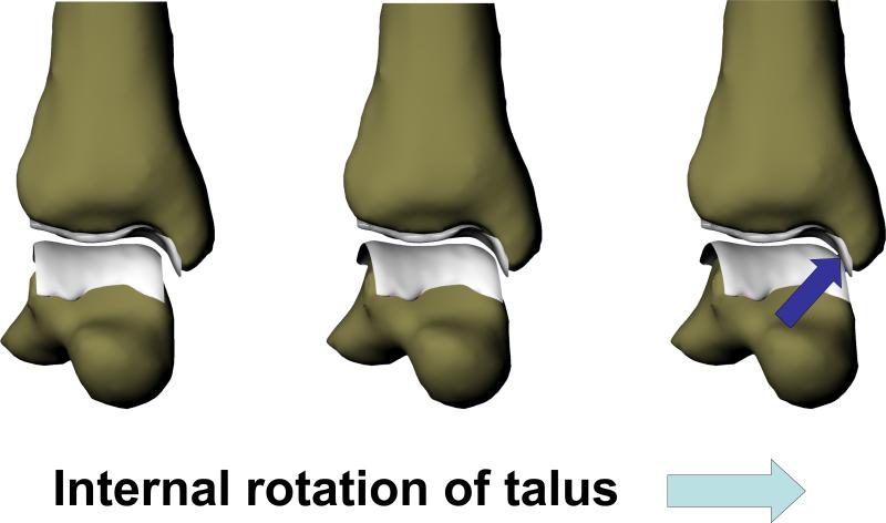
Hypothesis: Based on the orientation of the ATFL fibers, ATFL deficiency leads to increased anterior translation and increased internal rotation of the talus relative to the tibia.
Study design: Descriptive laboratory study.
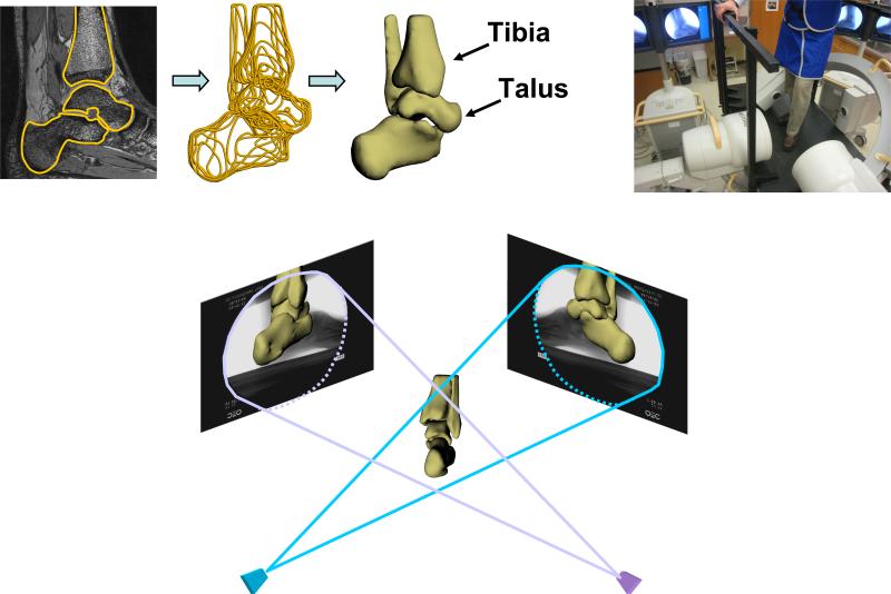
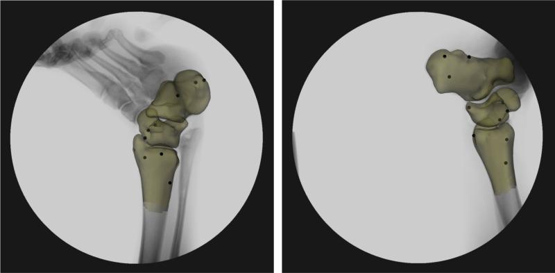
Methods: The ankles of 9 patients with unilateral ATFL injuries were compared as they stepped onto a level surface. Kinematic measurements were made as a function of increasing load. With use of magnetic resonance imaging and orthogonal fluoroscopy, the in vivo kinematics of the tibiotalar joint were measured in the ATFL-deficient and intact ankles of the same individuals.
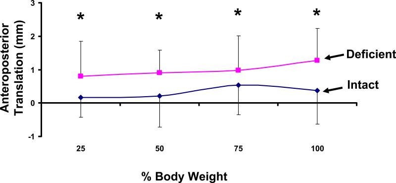
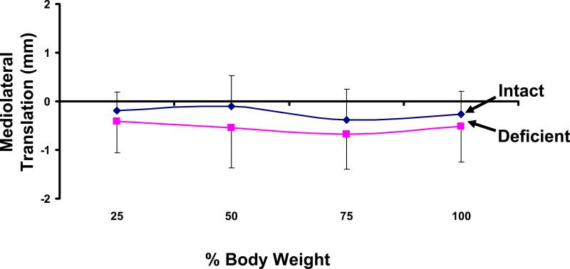
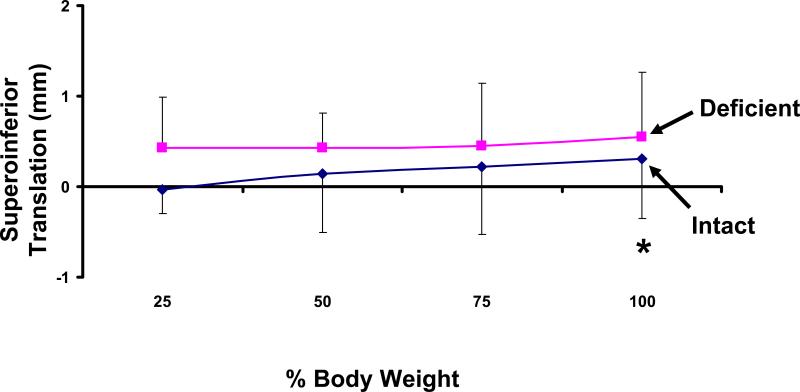
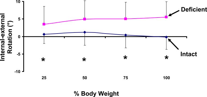
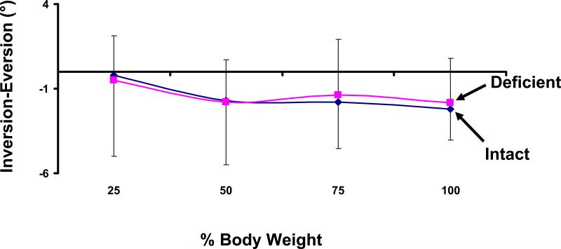
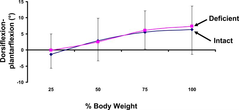
Results: A statistically significant increase in internal rotation, anterior translation, and superior translation of the talus was measured in ATFL-deficient ankles, as compared with the intact contralateral controls. For example, at 100% body weight, ATFL-deficient ankles demonstrated an increase of 0.9 +/- 0.5 mm in anterior translation (P = .008), an increase of 5.7 degrees +/- 3.6 degrees in internal rotation (P = .008), and a slight increase of 0.2 +/- 0.2 mm in the superior translation (P = .02) relative to the intact contralateral ankles.
Conclusion: Deficiency of the ATFL increases anterior translation, internal rotation, and superior translation of the talus.
Clinical relevance: Altered kinematics may contribute to the degenerative changes observed with chronic lateral ankle instability. These findings might help to explain the degenerative changes frequently observed on the medial talus in patients with chronic ATFL insufficiency and so provide a baseline for improving ankle ligament reconstructions aimed at restoring normal joint motion.
Figures
Similar articles
-
The effect of modified Broström-Gould repair for lateral ankle instability on in vivo tibiotalar kinematics.Am J Sports Med. 2012 Sep;40(9):2099-104. doi: 10.1177/0363546512454840. Epub 2012 Aug 10. Am J Sports Med. 2012. PMID: 22886690 Free PMC article.
-
Ankle stability in simulated lateral ankle ligament injuries.Foot Ankle Int. 2010 Jun;31(6):531-7. doi: 10.3113/FAI.2010.0531. Foot Ankle Int. 2010. PMID: 20557820
-
The Role of Calcaneofibular Ligament Injury in Ankle Instability: Implications for Surgical Management.Am J Sports Med. 2019 Feb;47(2):431-437. doi: 10.1177/0363546518815160. Epub 2018 Dec 20. Am J Sports Med. 2019. PMID: 30571138
-
Kinematics and Laxity of the Ankle Joint in Anatomic and Nonanatomic Anterior Talofibular Ligament Repair: A Biomechanical Cadaveric Study.Am J Sports Med. 2019 Mar;47(3):667-673. doi: 10.1177/0363546518820527. Epub 2019 Jan 25. Am J Sports Med. 2019. PMID: 30681886
-
The distal fascicle of the anterior inferior tibiofibular ligament as a cause of tibiotalar impingement syndrome: a current concepts review.Knee Surg Sports Traumatol Arthrosc. 2007 Apr;15(4):465-71. doi: 10.1007/s00167-006-0275-7. Epub 2007 Jan 20. Knee Surg Sports Traumatol Arthrosc. 2007. PMID: 17237964 Free PMC article. Review.
Cited by
-
Comparison of Broström technique, suture anchor repair, and tape augmentation for reconstruction of the anterior talofibular ligament.Knee Surg Sports Traumatol Arthrosc. 2016 Apr;24(4):1101-7. doi: 10.1007/s00167-015-3631-7. Epub 2015 May 10. Knee Surg Sports Traumatol Arthrosc. 2016. PMID: 25957613
-
Gait abnormalities in patients with chronic ankle instability can improve following a non-invasive biomechanical therapy: a retrospective analysis.J Phys Ther Sci. 2017 Apr;29(4):677-684. doi: 10.1589/jpts.29.677. Epub 2017 Apr 20. J Phys Ther Sci. 2017. PMID: 28533609 Free PMC article.
-
Healthy ankle and hindfoot kinematics during gait: Sex differences, asymmetry and coupled motion revealed through dynamic biplane radiography.J Biomech. 2021 Feb 12;116:110220. doi: 10.1016/j.jbiomech.2020.110220. Epub 2020 Dec 31. J Biomech. 2021. PMID: 33422727 Free PMC article.
-
Acute joint pathology and synovial inflammation is associated with increased intra-articular fracture severity in the mouse knee.Osteoarthritis Cartilage. 2011 Jul;19(7):864-73. doi: 10.1016/j.joca.2011.04.011. Epub 2011 May 12. Osteoarthritis Cartilage. 2011. PMID: 21619936 Free PMC article.
-
Recommendation of minimal distal tibial length for long axis coordinate system definitions.J Biomech. 2024 Jun;170:112153. doi: 10.1016/j.jbiomech.2024.112153. Epub 2024 May 16. J Biomech. 2024. PMID: 38795543 Free PMC article.
References
-
- Ajis A, Younger AS, Maffulli N. Anatomic repair for chronic lateral ankle instability. Foot Ankle Clin. 2006 Sep;11(3):539–545. - PubMed
-
- Andriacchi TP, Briant PL, Bevill SL, Koo S. Rotational changes at the knee after ACL injury cause cartilage thinning. Clin Orthop Relat Res. 2006 Jan;442:39–44. - PubMed
-
- Andriacchi TP, Mundermann A, Smith RL, Alexander EJ, Dyrby CO, Koo S. A framework for the in vivo pathomechanics of osteoarthritis at the knee. Ann Biomed Eng. 2004 Mar;32(3):447–457. - PubMed
-
- Aydogan U, Glisson RR, Nunley JA. Extensor retinaculum augmentation reinforces anterior talofibular ligament repair. Clin Orthop Relat Res. 2006 Jan;442:210–215. - PubMed
Publication types
MeSH terms
Grants and funding
LinkOut - more resources
Full Text Sources
Other Literature Sources
Medical
