. 2010 Jan;51(1):150-61.
doi: 10.1194/M900346-JLR200.

Expression of apolipoprotein C-III in McA-RH7777 cells enhances VLDL assembly and secretion under lipid-rich conditions

Affiliations

Expression of apolipoprotein C-III in McA-RH7777 cells enhances VLDL assembly and secretion under lipid-rich conditions

Meenakshi Sundaram et al. J Lipid Res. 2010 Jan.

Abstract

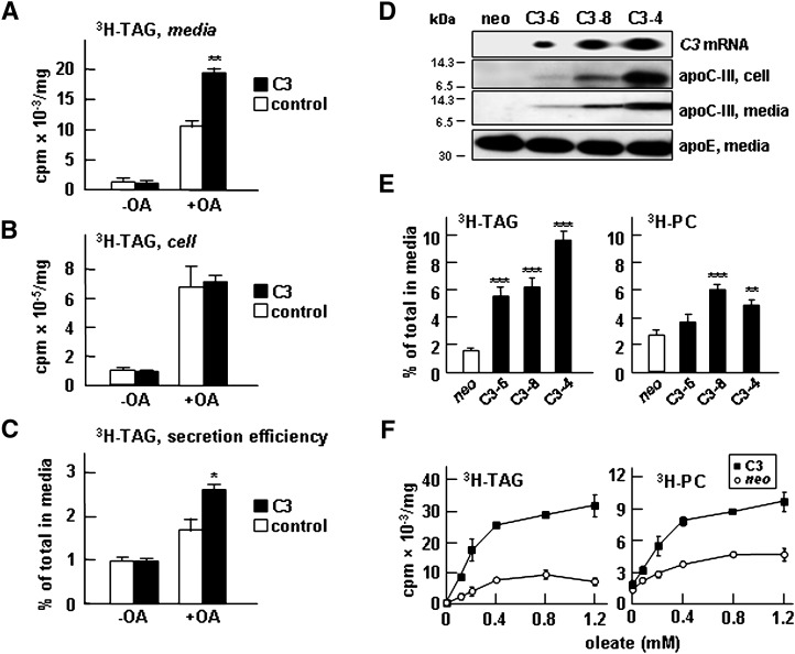
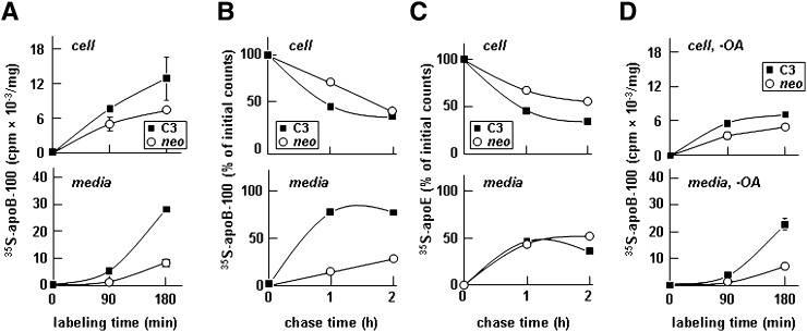
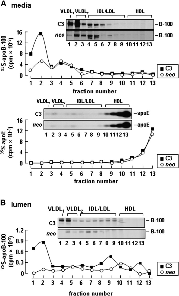
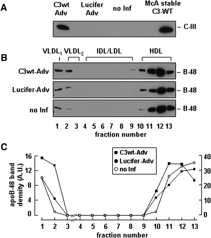
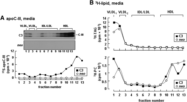
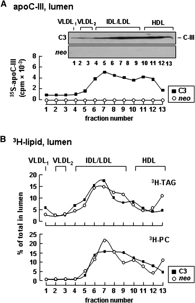
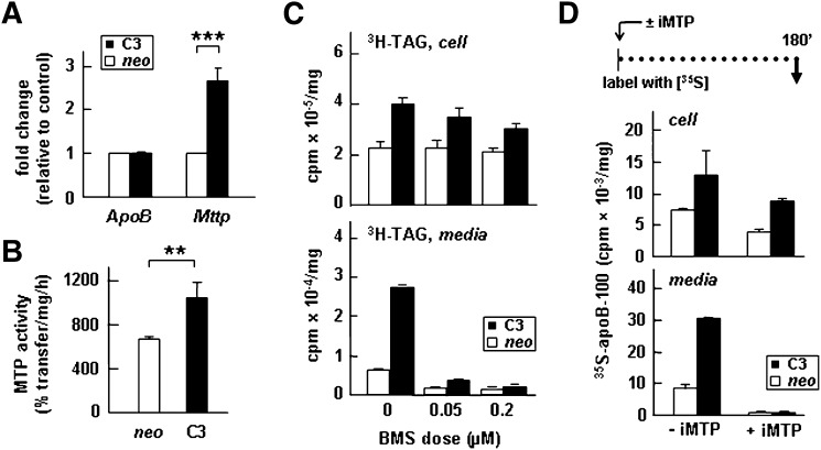
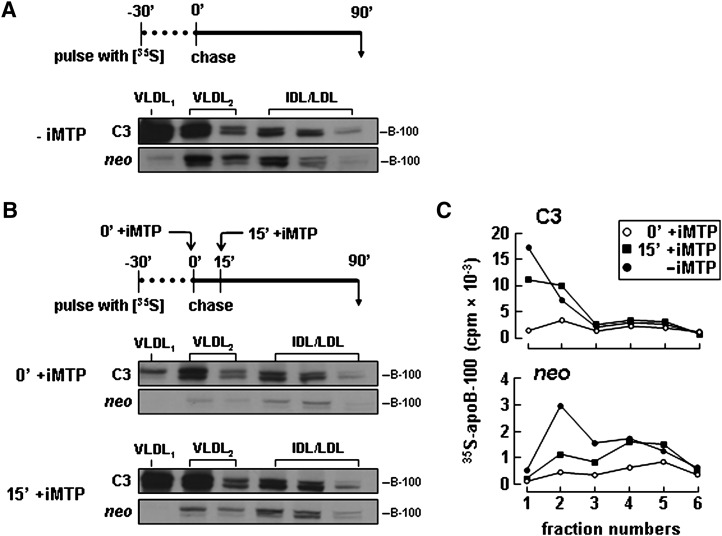
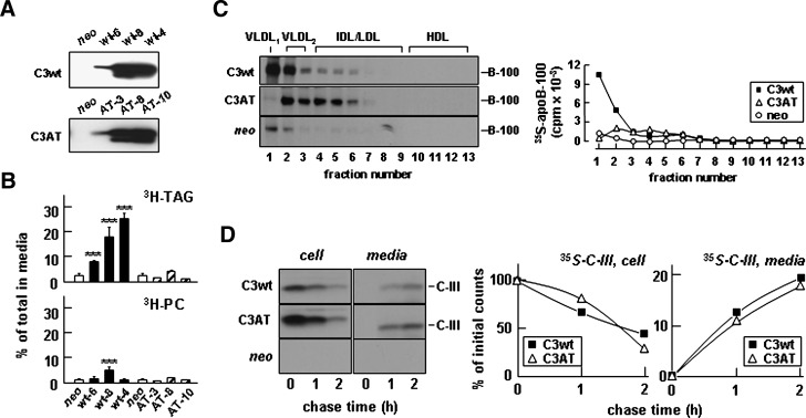
Apolipoprotein (apo) C-III plays a regulatory role in VLDL lipolysis and clearance. In this study, we determined a potential intracellular role of apoC-III in hepatic VLDL assembly and secretion. Stable expression of recombinant apoC-III in McA-RH7777 cells resulted in increased secretion efficiency of VLDL-associated triacylglycerol (TAG) and apoB-100 in a gene-dosage-dependent manner. The stimulatory effect of apoC-III on TAG secretion was manifested only when cells were cultured under lipid-rich (i.e., media supplemented with exogenous oleate) but not lipid-poor conditions. The stimulated TAG secretion was accompanied by increased secretion of apoB-100 and apoB-48 as VLDL(1). Expression of apoC-III also increased mRNA and activity of microsomal triglyceride transfer protein (MTP). Pulse-chase experiments showed that apoC-III expression promoted VLDL(1) secretion even under conditions where the MTP activity was inhibited immediately after the formation of lipid-poor apoB-100 particles, suggesting an involvement of apoC-III in the second-step VLDL assembly process. Consistent with this notion, the newly synthesized apoC-III was predominantly associated with TAG within the microsomal lumen that resembled lipid precursors of VLDL. Introducing an Ala23-to-Thr mutation into apoC-III, a naturally occurring mutation originally identified in two Mayan Indian subjects with hypotriglyceridemia, abolished the ability of apoC-III to stimulate VLDL secretion from transfected cells. Thus, expression of apoC-III in McA-RH7777 cells enhances hepatic TAG-rich VLDL assembly and secretion under lipid-rich conditions.


Figures

Fig. 1.
Fig. 1.
Expression of apoC-III in McA-RH7777 cells resulted in enhanced [3H]TAG secretion under lipid-rich conditions. Control or transiently apoC-III (C3) transfected cells (48 h posttransfection) were labeled with [3H]glycerol for 2 h in DMEM containing 20% FBS ± 0.4 mM oleate. At the end of labeling, radioactivity associated with [3H]TAG in media (A) and cells (B) was quantified. C: [3H]TAG secretion efficiency is expressed as percentage of total [3H]TAG (cell + media) that was secreted into media. D: Characterization of three stable clones (C3-6, C3-8, and C3-4) expressing different levels of apoC-III. From top to bottom, Northern blots for APOC3 mRNA, immunoblots for apoC-III in cells, immunoblots for apoC-III in media, and immunoblots for endogenous apoE. E: Secretion efficiency of [3H]TAG and [3H]PC from the stable apoC-III clones. The indicated cells were labeled with [3H]glycerol for 4 h in DMEM containing 20% FBS + 0.4 mM oleate. Statistical significance ***P < 0.001; **P < 0.01; *P < 0.05 (Student's t-test of C3 versus control). Error bars indicate ± SD (n = 3). F: Control (neo) and cells expressing apoC-III (C3) were labeled with [3H]glycerol for 2 h in DMEM containing 20% FBS and increasing concentrations of oleate. At the end of labeling, radioactivity associated with [3H]TAG (left panel) and [3H]PC (right panel) in media and cells was quantified.
Fig. 2.
Fig. 2.
Expression of apoC-III-enhanced secretion of [35S]apolipoprotein B-100 (apoB-100). A: Control (neo) and apoC-III-expressing cells (C3) were continuously labeled with [35S]methionine/cysteine for 90 or 180 min in DMEM containing 20% FBS + 0.4 mM oleate. B and C: Cells were pulse labeled for 30 min and “chased” for up to 2 h in DMEM containing 20% FBS + 0.4 mM oleic acid (OA). D: Cells were continuously labeled for 90 or 180 min in the media without OA. At the indicated times, [35S]apoB-100 (A, B, and D) and [35S]apoE (C) were recovered from the cells and media by immunoprecipitation, resolved by SDS-PAGE, and subjected to fluorography. Radioactivity associated with [35S]apoB and [35S]apoE in cells and media was quantified. For the continuous labeling experiments (A and D), data are presented as cpm/mg cell protein. For the pulse-chase experiments (B and C), data are presented as percentage of initial counts, which are the counts associated with cell [35S]apoB-100 or cell [35S]apoE at the end of a 30 min pulse. The experiment was repeated, and similar results were obtained.
Fig. 3.
Fig. 3.
Expression of apoC-III increased [35S]apolipoprotein B-100 (apoB-100) secretion as VLDL1 and VLDL2. A: Representative fluorograms and quantification of [35S]apoB-100 (top panel) and [35S]apoE (bottom panel) distribution among medium lipoproteins. Control (neo) or apoC-III expressing cells (C3) were labeled with [35S]methionine/cysteine for 3 h in the presence of 20% serum + 0.4 mM oleate. After labeling, the media were subjected to cumulative rate flotation ultracentrifugation. The apoB-100 and apoE proteins associated with each fraction were recovered by immunoprecipitation, resolved by SDS-PAGE, and visualized by fluorography. Radioactivity associated with [35S]apoB-100 and apoE was quantified by scintillation counting. B: Representative fluorograms and quantification of [35S]apoB-100 distribution among lipoproteins within the microsomal lumen. The cells were labeled with [35S]methionine/cysteine for 60 min in the presence of 20% serum + 0.4 mM oleate. The content of microsomal lumen was fractionated by ultracentrifugation, [35S]apoB-100 in each fraction was analyzed by SDS-PAGE, and the associated radioactivity was quantified as described above. The experiment was repeated, and similar results were obtained.
Fig. 4.
Fig. 4.
Adenovirus-mediated apoC-III expression stimulated apoB-48 secretion as VLDL. McA-RH7777 cells stably expressing human apoB-48 ( 35 ) were infected with adenovirus vectors encoding C3-wt or luciferase genes for 4 h and cultured for additional 48 h in DMEM supplemented with 20% serum. A: 48 h after infection, the cells were cultured in the same media + 0.4 mM OA for 8 h, and the conditioned media were collected to determine apoC-III secretion. B: The conditioned media were fractioned by cumulative rate flotation, and the apoB-48 proteins were recovered from each fraction by immunoprecipitation, resolved by SDS-PAGE, and detected by immunoblotting. C: Scanning densitometry of apo-B48 bands. (Note the scale for VLDL fractions is enlarged.)
Fig. 5.
Fig. 5.
Expression of apoC-III increased secretion as VLDL containing [35S]apoC-III and [3H]TAG. A: Control (neo) or apoC-III-expressing cells (C3) were labeled with either [35S]methionine/cysteine for 3 h or [3H]glycerol for 4 h in DMEM containing 20% serum + 0.4 mM oleate. The conditioned media were fractionated by rate flotation ultracentrifugation, and metabolic labeled proteins and lipids were recovered from each fraction. A: Distribution of [35S]apoC-III among the fractionated lipoproteins. B: Distribution of [3H]TAG (top panel) and [3H]PC (bottom panel). The experiment was repeated, and similar results were obtained.
Fig. 6.
Fig. 6.
Distribution of apoC-III and TAG among lipoproteins within the microsomal lumen. Control (neo) or apoC-III-expressing cells (C3) were labeled with [35S]methionine/cysteine for 60 min or with [3H]glycerol for 60 min in DMEM containing 20% serum + 0.4 mM oleate. The content of microsomal lumen was fractionated by ultracentrifugation as described in Fig. 3B. A: Distribution of [35S]apoC-III among lipoproteins within the microsomal lumen. B: Distribution of [3H]TAG (top panel) and [3H]PC (bottom panel). The [3H]TAG and [3H]PC in each fraction is expressed as percentage of total lumenal [3H]TAG or [3H]PC.
Fig. 7.
Fig. 7.
Expression of apoC-III increased the expression of Mttp gene and MTP activity. A: The relative mRNA concentrations of apoB and MTP (with respect to 18s rRNA) were quantified by real-time RT-PCR. The data are presented as fold changes between apoC-III-expressing cells (C3) and control (neo). B: MTP activity assay. Data are the average of triplicate assays using cell lysate obtained from three different stable apoC-III-expressing clones (C3) or neo controls. ***P < 0.001; **P < 0.01 (Student's t-test of C3 versus control). Error bars indicate ± SD (n = 3). C: Cells were labeled with [3H]glycerol for 3 h as in Fig. 1E in the absence or presence of 0.05 or 0.2 µM BMS-197636 (BMS). At the end of labeling, radioactivity associated with [3H]TAG in media and cells was quantified as described in Fig. 1A, B. D: Control (neo) and apoC-III-expressing cells (C3) were continuously labeled with [35S]methionine/cysteine for 180 min in the presence or absence of 0.2 µM BMS-197636 (± iMTP). The labeling media were supplemented with 0.4 mM OA. The apoB-100 protein was recovered from the cell (top panel) and media (bottom panel) by immunoprecipitation, resolved by SDS-PAGE, and followed by fluorography. Radioactivity associated with [35S]apoB-100 was quantified by scintillation counting.
Fig. 8.
Fig. 8.
Secretion of apoB-100 as VLDL1 from apoC-III-expressing cells was less sensitive to MTP inactivation. A: Cells were pulse labeled for 30 min with [35S]methionine/cysteine in media supplemented with 0.4 mM OA in the absence of MTP inhibitor (-iMTP) and then “chased” for 90 min with DMEM supplemented with 20% FBS and 0.4 mM OA in the absence of MTP inhibitor (-iMTP). B: Cells were pulse-labeled for 30 min as in A. During chase, the MTP inhibitor (+ iMTP) was included either at the beginning of chase (0′+ iMTP) or 15 min after (15′+ iMTP) the start of chase. The media were collected at the end of 90 min chase and subjected to cumulative rate floatation centrifugation. The apoB-100 protein associated with each fraction was recovered by immunoprecipitation, resolved by SDS-PAGE, and visualized by fluorography. C: Plot of the radioactivity associated with [35S]apoB-100 shown in A and B was quantified by scintillation counting. (Note that only fractions 1 through 6 that contained apoB-100 are presented.)
Fig. 9.
Fig. 9.
The Ala23Thr mutation compromised the ability of apoC-III to stimulate VLDL secretion. A: Immunoblots of apoC-III (C3wt) or Ala23Thr mutant (C3AT) secreted from different stable cell lines. B: Secretion efficiency of [3H]TAG (top panel) and [3H]PC (bottom panel). The experiments were performed as described in the legend of Fig. 1E. C: Representative fluorograms (left panel) and quantification of [35S]apoB-100 (right panel) associated with fractionated medium lipoproteins. The experiment was performed essentially the same as described in the legend of Fig. 3A, except cells expressing C3wt or the C3AT mutant were used. D: Secretion efficiency of apoC-III. The cells were pulse-labeled with [35S]methionine/cysteine for 30 min and “chased” for up to 2 h. [35S]apoC-III was recovered from cell and media, respectively, resolved by SDS-PAGE, and visualized by fluorography. Left panel, fluorograms of cell and medium-associated [35S]apoC-III. Right panel, radioactivity associated with [35S]apoC-III was quantified by scintillation counting. Data are expressed as percentage of initial [35S]apoC-III associated with cell or secreted into media during chase. ***P < 0.001 (Student's t-test of C3wt versus control). Error bars indicate ± SD (n = 3).

Similar articles

Cited by

References

    1. Jong M. C., Hofker M. H., Havekes L. M. 1999. Role of ApoCs in lipoprotein metabolism: functional differences between ApoC1, ApoC2, and ApoC3. Arterioscler. Thromb. Vasc. Biol. 19: 472–484. - PubMed
    1. Cohn J. S., Tremblay M., Batal R., Jacques H., Rodriguez C., Steiner G., Mamer O., Davignon J. 2004. Increased apoC-III production is a characteristic feature of patients with hypertriglyceridemia. Atherosclerosis. 177: 137–145. - PubMed
    1. Zheng C., Khoo C., Ikewaki K., Sacks F. M. 2007. Rapid turnover of apolipoprotein C–III-containing triglyceride-rich lipoproteins contributing to the formation of LDL subfractions. J. Lipid Res. 48: 1190–1203. - PubMed
    1. Karathanasis S. K., Norum R. A., Zannis V. I., Breslow J. L. 1983. An inherited polymorphism in the human apolipoprotein A-I gene locus related to the development of atherosclerosis. Nature. 301: 718–720. - PubMed
    1. Karathanasis S. K., Ferris E., Haddad I. A. 1987. DNA inversion within the apolipoproteins AI/CIII/AIV-encoding gene cluster of certain patients with premature atherosclerosis. Proc. Natl. Acad. Sci. USA. 84: 7198–7202. - PMC - PubMed

Publication types