. 2009 Aug;65(Pt 8):786-95.
doi: 10.1107/S0907444909015935. Epub 2009 Jul 10.

Adaptability and selectivity of human peroxisome proliferator-activated receptor (PPAR) pan agonists revealed from crystal structures

Affiliations

Adaptability and selectivity of human peroxisome proliferator-activated receptor (PPAR) pan agonists revealed from crystal structures

Takuji Oyama et al. Acta Crystallogr D Biol Crystallogr. 2009 Aug.

Abstract

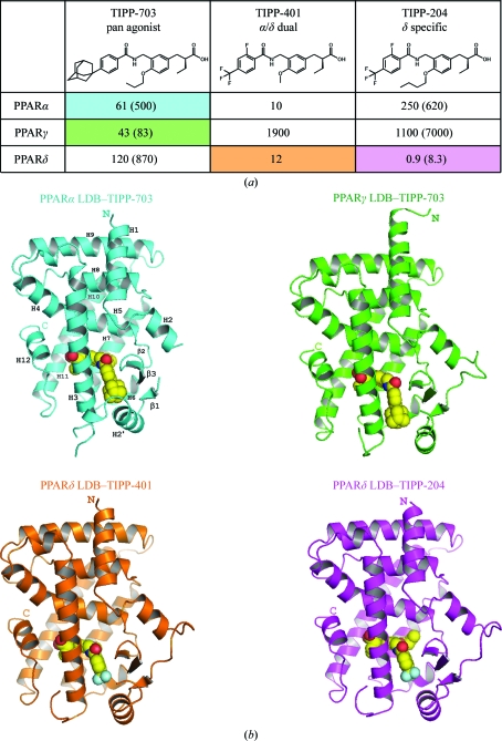
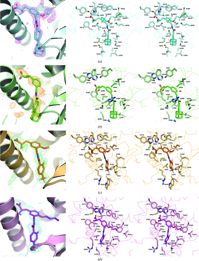
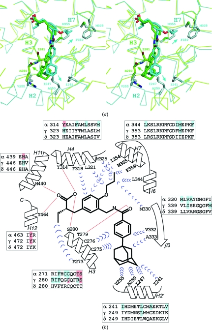
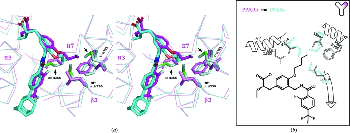
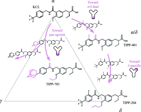
Peroxisome proliferator-activated receptors (PPARs) belong to the nuclear hormone receptor family, which is defined as transcriptional factors that are activated by the binding of ligands to their ligand-binding domains (LBDs). Although the three PPAR subtypes display different tissue distribution patterns and distinct pharmacological profiles, they all are essentially related to fatty-acid and glucose metabolism. Since the PPARs share similar three-dimensional structures within the LBDs, synthetic ligands which simultaneously activate two or all of the PPARs could be potent candidates in terms of drugs for the treatment of abnormal metabolic homeostasis. The structures of several PPAR LBDs were determined in complex with synthetic ligands, derivatives of 3-(4-alkoxyphenyl)propanoic acid, which exhibit unique agonistic activities. The PPARalpha and PPARgamma LBDs were complexed with the same pan agonist, TIPP-703, which activates all three PPARs and their crystal structures were determined. The two LBD-ligand complex structures revealed how the pan agonist is adapted to the similar, but significantly different, ligand-binding pockets of the PPARs. The structures of the PPARdelta LBD in complex with an alpha/delta-selective ligand, TIPP-401, and with a related delta-specific ligand, TIPP-204, were also determined. The comparison between the two PPARdelta complexes revealed how each ligand exhibits either a ;dual selective' or ;single specific' binding mode.


Figures

Figure 1
Figure 1
Crystal structures of PPAR LBD–TIPP complexes. (a) Chemical formulae of TIPP-703, TIPP-401 and TIPP-204. The numbers indicate the EC50 (nM), the molar concentration of the compounds that affords 50% of the maximal reporter activity, in our PPAR-GAL4 chimeric reporter assays using transiently transfected HEK-293 cells (Kasuga et al., 2006 ▶). Values in parentheses indicate the activities of the antipodal (R) isomers of TIPP-703 and TIPP-401. For the structures of the four complexes determined in this study, the columns in the table are coloured cyan (PPARα LBD–TIPP-703), green (PPARγ LBD–TIPP-703), orange (PPARδ LBD–TIPP-401) and magenta (PPARδ LBD–TIPP-204). This colouring is used throughout the manuscript. (b) Overall structures of the complexes. Proteins are represented as ribbon models and the ligands are depicted as space-filling models, with F, C, N and O atoms in aqua, yellow, blue and red, respectively.
Figure 2
Figure 2
Close-up views of the ligand-binding pockets. (a) PPARα LBD–TIPP-703. (b) PPARγ LBD–TIPP-703. (c) PPARδ LBD–TIPP-401. (d) PPARδ LBD–TIPP-204. The left column shows the OMIT F oF c electron-density maps (contoured at 2.2σ) and the right columns show stereoviews of the interaction between the ligand-binding pockets and the bound ligands. The amino-acid residues contacting the ligands are labelled.
Figure 3
Figure 3
Comparison between the PPARα LBD–TIPP-703 and the PPARγ LBD–TIPP-703 complexes. (a) Stereoview of the superimposed structures. The PPARα LBD–TIPP-703 complex is coloured cyan and the PPARγ LBD–TIPP-703 complex is coloured green. The key contact residues in the complexes are highlighted. Prominent interactions in each complex are indicated by arrows. (b) Schematic view of the protein–ligand interactions. A structure-based sequence alignment was generated around the ligand-binding pocket. In the PPARα and PPARγ sequences, the residues that interact with TIPP-703 are coloured blue (hydrophobic) and red (hydrophilic).
Figure 4
Figure 4
Comparison between the PPARα LBD–TIPP-703 and the PPARδ LBD–TIPP-204 complexes. (a) Stereoview of the superimposed structures. The PPARα LBD–TIPP-703 complex is coloured cyan and the PPARδ LBD–TIPP-204 complex is coloured magenta. The key contact residues of the complexes are highlighted. Prominent interactions in each complex are indicated by arrows. (b) Schematic view of the protein–ligand interactions, highlighting the specificity of TIPP-204 toward PPARδ.
Figure 5
Figure 5
Summary of the development of the present TIPP compounds from a PPARα-specific agonist, KCL. The TIPP agonists used in this study were all developed from KCL using structure–activity relation (SAR) studies. When specific chemical groups, coloured magenta, were introduced into the ligand compounds, the transactivation abilities were drastically changed. The modification effects of the agonist ligands were found to increase the protein–ligand interactions at specific positions in the ligand-binding pockets.

References

    1. Artis, D. R. et al. (2009). Proc. Natl Acad. Sci. USA, 106, 262–267. - PubMed
    1. Banner, C. D., Gottlicher, M., Widmark, E., Sjövall, J., Rafter, J. J. & Gustafsson, J. A. (1993). J. Lipid Res.34, 1583–1591. - PubMed
    1. Brünger, A. T., Adams, P. D., Clore, G. M., DeLano, W. L., Gros, P., Grosse-Kunstleve, R. W., Jiang, J.-S., Kuszewski, J., Nilges, M., Pannu, N. S., Read, R. J., Rice, L. M., Simonson, T. & Warren, G. L. (1998). Acta Cryst. D54, 905–921. - PubMed
    1. Bruning, J. B., Chalmers, M. J., Prasad, S., Busby, S. A., Kamenecka, T. M., He, Y., Nettles, K. W. & Griffin, P. R. (2007). Structure, 15, 1258–1271. - PubMed
    1. Casimiro-Garcia, A. et al. (2008). Bioorg. Med. Chem.16, 4883–4907. - PubMed

Publication types

MeSH terms