. 2009 Oct;17(5):381-7.
doi: 10.1097/MOO.0b013e3283303347.

Regulation of cell fate and patterning in the developing mammalian cochlea

Affiliations

Regulation of cell fate and patterning in the developing mammalian cochlea

Matthew W Kelley et al. Curr Opin Otolaryngol Head Neck Surg. 2009 Oct.

Abstract

Purpose of review: A significant proportion of hearing loss and deafness is caused by defects in the structure or function of cells within the organ of Corti. Identification of the molecular factors that regulate the development of this structure should provide valuable insights regarding inner ear formation and the signaling pathways that underlie congenital auditory deficits. In addition, targeted modulation of these same factors could be developed as therapies for hair cell regeneration.

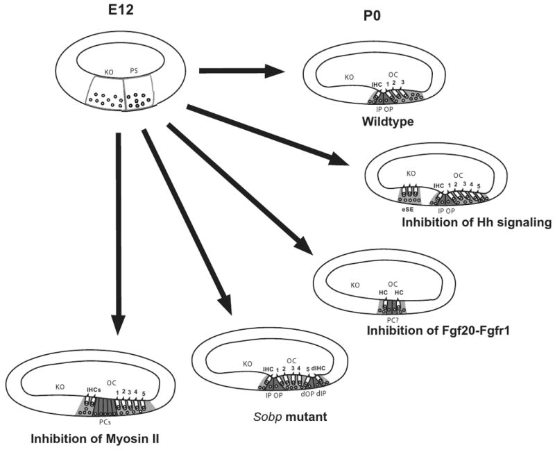
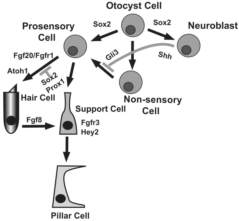
Recent findings: Results from experiments using transgenic and mutant mice, as well as in-vitro techniques, have identified genes and signaling pathways that are required to either specify unique auditory cell types, such as hair cells or supporting cells, or to generate the highly ordered cellular pattern that is characteristic for the organ of Corti. In particular, the hedgehog and fibroblast growth factor signaling pathways modulate the formation of the progenitor cells that will give rise to the organ of Corti. SRY-box containing gene 2, a transcription factor that is required for the formation of the cochlear progenitor cell population, has paradoxically been shown to also act as an inhibitor of hair cell development. Finally, the motor protein myosin II regulates extension of the organ of Corti and the alignment of hair cells and supporting cells into ordered rows.

Summary: A better understanding of the signaling pathways that direct different aspects of cochlear development, such as specific of cell fates or cellular patterning, offers the potential to identify new pathways or molecules that could be targeted for therapeutic interventions.


Figures

Figure 1
Figure 1
Phenotypic changes in the organ of Corti in response to changes in specific signaling pathways. At E12, the floor of the developing cochlear duct is comprised of undifferentiated cells that can be classified as part of the prosensory domain (PS) or Kolliker's organ (KO) based on relative position. By P0, the normal cellular pattern of the organ of Corti (OC), including a single inner hair cell (IHC), three outer hair cells (numbered), inner and outer pillar cells (IP and OP) and supporting cells (light gray), is present. Inhibition of hedgehog signaling leads to the formation of ectopic hair cells and supporting cells (eSE) in Kolliker's organ as well as an overproduction of outer hair cells. In contrast, inhibition of Fgf20-Fgfr1 interactions leads to the formation of small patches of unpatterned hair cells and supporting cells including some cells that may be pillar cells. In Sobp mutants, mirror-image duplications of the organ of Corti are present including a duplicated inner hair cells (dIHC) and duplicated pillar cells (dIP and dOP). Finally, inhibition of Myosin II leads to defects in extension of the cochlear duct. In a cross-section this appears as additional rows of all cell types.
Figure 2
Figure 2
Lineage relationships of cells originating in the otocyst. Cells within the otocyst have three broad fates, Prosensory Cell, Non-sensory cell, or Neuroblast. As discussed in the text, Sox2 actually plays a role in the specification of both the Prosensory and Neuroblast lineages. Prosensory cells then make a secondary choice between Hair Cell and Supporting Cell fates, with activation of Fgfr1 by Fgf20 positively influencing the hair cell fate, leading to activation of Atoh1. In contrast, Sox2 acts as an inhibitor of the hair cell fate at this decision point, probably at least in some cells through activation of Prox1. For the particular case of pillar cells, developing inner hair cells express Fgf8 which activates Fgfr3 in adjacent progenitors, leading to an inhibition of the hair cell fate and an induction of the pillar cell fate in those cells. Finally, the hedgehog signaling pathway acts to prevent at least some Non-Sensory Cells from spontaneously switching into the Prosensory lineage.

References

    1. Cotanche DA. Genetic and pharmacological intervention for treatment/prevention of hearing loss. J Commun Disord. 2008;41:421–443. - PMC - PubMed
    1. Collado MS, Burns JC, Hu Z, Corwin JT. Recent advances in hair cell regeneration research. Curr Opin Otolaryngol Head Neck Surg. 2008;16:465–471. - PMC - PubMed
    1. Kikuchi T, Adams JC, Miyabe Y, So E, Kobayashi T. Potassium ion recycling pathway via gap junction systems in the mammalian cochlea and its interruption in hereditary nonsyndromic deafness. Med Electron Microsc. 2000;33:51–56. - PubMed
    1. Barald KF, Kelley MW. From placode to polarization: new tunes in inner ear development. Development. 2004;131:4119–4130. - PubMed
    1. Kelley MW. Regulation of cell fate in the sensory epithelia of the inner ear. Nat Rev Neurosci. 2006;7:837–849. - PubMed

Publication types

MeSH terms