. 2009 Aug 18;101(4):666-72.
doi: 10.1038/sj.bjc.6605196. Epub 2009 Jul 21.

Angiogenesis is associated with the onset of hyperplasia in human ductal breast disease

Affiliations

Angiogenesis is associated with the onset of hyperplasia in human ductal breast disease

J E Bluff et al. Br J Cancer. .

Abstract

Background: The precise timing of the angiogenic switch and the role of angiogenesis in the development of breast malignancy is currently unknown.

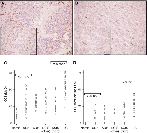
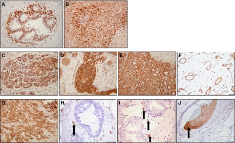
Methods: Therefore, the expression of CD31 (pan endothelial cells (ECs)), endoglin (actively proliferating ECs), hypoxia-inducible factor-1 (HIF-1alpha), vascular endothelial growth factor-A (VEGF) and tissue factor (TF) were quantified in 140 surgical specimens comprising normal human breast, benign and pre-malignant hyperplastic tissue, in situ and invasive breast cancer specimens.

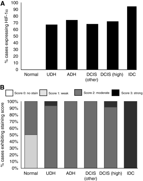
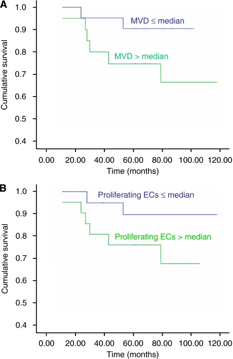
Results: Significant increases in angiogenesis (microvessel density) were observed between normal and benign hyperplastic breast tissue (P<0.005), and between in situ and invasive carcinomas (P<0.0005). In addition, significant increases in proliferating ECs were observed in benign hyperplastic breast compared with normal breast (P<0.05) cancers and in invasive compared with in situ cancers (P<0.005). Hypoxia-inducible factor-1alpha, VEGF and TF expression were significantly associated with increases in both angiogenesis and proliferating ECs (P<0.05). Moreover, HIF-1alpha was expressed by 60-75% of the hyperplastic lesions, and a significant association was observed between VEGF and TF in ECs (P<0.005) and invasive tumour cells (P<0.01).

Conclusions: These findings are the first to suggest that the angiogenic switch, associated with increases in HIF-1alpha, VEGF and TF expression, occurs at the onset of hyperplasia in the mammary duct, although the greatest increase in angiogenesis occurs with the development of invasion.


Figures

Figure 1
Figure 1
Endothelial staining in breast lesions. Immunohistochemical staining of ECs with (A) CD31 and (B) endoglin in high-grade DCIS. Note the increased frequency and intensity of stained ECs for CD31 compared with endoglin (scale=50 μm). Scatter plots representing cumulative Chalkley scores in different breast tissue samples immunostained with (C) CD31 (MVD) and (D) endoglin (proliferating ECs). (C) There was a significant increase in MVD between normal breast tissue and UDH (P<0.005), and a further significant increase in MVD between in situ (DCIS) and invasive breast cancers (P<0.0005). (D) There was a significant increase in proliferating ECs in UDH cases compared with normal breast (P<0.05) and in IDC compared with high-grade DCIS specimens (P<0.005). P<0.05 was considered significant (Kruskal–Wallis followed by Mann–Whitney U-test).
Figure 2
Figure 2
Immunohistochemical staining for HIF-1α, VEGF and TF. (A) Nuclear HIF-1α staining in the ductal epithelial cells of an ADH case and (B) tumour cells of an invasive cancer. (C) Weak expression of VEGF in normal breast epithelium (score=1), (D) strong expression in florid usual ductal hyperplasia (score=2/3) and (E) strong staining localised to tumour cells within invasive breast carcinomas (score=3). (F) VEGF expression in ECs in normal breast tissue. (G) Tumour cells expressed TF in approximately 55% of invasive breast cancer specimens. (H) TF was expressed in ECs associated with benign hyperplastic tissue (arrow). (I) Putative macrophages expressing TF associated with areas of DCIS (arrows). (J) TF expressed in vessel containing thrombosis (arrow). Photographs AE and G were taken at × 20 magnification and all others at × 40 magnification.
Figure 3
Figure 3
Expression of HIF-1α and VEGF in the epithelial/tumour cells of breast lesions. (A) Percentage of cases expressing HIF-1α. There is a significant increase in HIF-1α expression seen with increasing severity of lesion (P<0.01) (B) Percentage of cases with varying VEGF expression. There is a significant increase in VEGF expression seen with increasing severity of lesion (P<0.001).
Figure 4
Figure 4
Cox regression survival graphs. Comparison of overall survival between groups of patients with ⩽median and >median cumulative Chalkley scores (CCS) for (A) MVD (CD31; P=0.09) and (B) proliferating ECs (endoglin; P=0.12).

Similar articles

Cited by

References

    1. Abe K, Shoji M, Chen J, Bierhaus A, Danave I, Micko C, Casper K, Dillehay DL, Nawroth PP, Rickles FR (1999) Regulation of vascular endothelial growth factor production and angiogenesis by the cytoplasmic tail of tissue factor. Proc Natl Acad Sci USA 96: 8663–8668 - PMC - PubMed
    1. Amarzguioui M, Peng Q, Wiiger MT, Vasovic V, Babaie E, Holen T, Nesland JM, Prydz H (2006) Ex vivo and in vivo delivery of anti-tissue factor short interfering RNA inhibits mouse pulmonary metastasis of B16 melanoma cells. Clin Cancer Res 12: 4055–4061 - PubMed
    1. Armesilla AL, Lorenzo E, Gómez de Arco P, Martinez-Martinez S, Alfranca A, Redondo JM (1999) Vascular endothelial growth factor activates nuclear factor of activated T cells in human endothelial cells: a role for tissue factor gene expression. Mol-Cell Biol 19: 2032–2043 - PMC - PubMed
    1. Beresford MJ, Harris AL, Ah-See M, Daley F, Padhani AR, Makris A (2006) The relationship of the neo-angiogenic marker, endoglin, with response to neoadjuvant chemotherapy in breast cancer. Br J Cancer 95: 1683–1688 - PMC - PubMed
    1. Bergers G, Benjamin LE (2003) Tumorigenesis and the angiogenic switch. Nat Rev Cancer 3: 401–410 - PubMed

Publication types

MeSH terms

Substances