. 2009 Jun 11:3:2.
doi: 10.3389/neuro.03.002.2009. eCollection 2009.

Drosophila photoreceptors and signaling mechanisms

Affiliations

Drosophila photoreceptors and signaling mechanisms

Ben Katz et al. Front Cell Neurosci. .

Abstract

Fly eyes have been a useful biological system in which fundamental principles of sensory signaling have been elucidated. The physiological optics of the fly compound eye, which was discovered in the Musca, Calliphora and Drosophila flies, has been widely exploited in pioneering genetic and developmental studies. The detailed photochemical cycle of bistable photopigments has been elucidated in Drosophila using the genetic approach. Studies of Drosophila phototransduction using the genetic approach have led to the discovery of novel proteins crucial to many biological processes. A notable example is the discovery of the inactivation no afterpotential D scaffold protein, which binds the light-activated channel, its activator the phospholipase C and it regulator protein kinase C. An additional protein discovered in the Drosophila eye is the light-activated channel transient receptor potential (TRP), the founding member of the diverse and widely spread TRP channel superfamily. The fly eye has thus played a major role in the molecular identification of processes and proteins with prime importance.

Keywords: G-protein; INAD scaffold protein; TRP channels; bistable pigments; optics of compound eyes; phosphoinositide cycle; phospholipase C; phosphorylated arrestin.


Figures

Figure 1
Figure 1
The morphology and optics of the compound eye. (A) The compound eye of Musca and the visual ganglionic layers: a schematic representation of a horizontal section. Inset – Schematic representation of the distal area of a single ommatidium. C – corneal lens, PC – pseudocone, RZ – retinula cells (photoreceptor), PZ – pigment cells, K – rhabdomere cap, SZ – Semper cells, Rh – rhabdomere, La – lamina, Me – medulla (modified from Kirschfeld, 1967). (B,C) Electron microscopic (EM) cross-section of Drosophila ommatidia and a rhabdomere at the upper region of the photoreceptors respectively. M – microvilli, SMC – submicrovillar cisternae, N – nucleus (modified from Minke and Selinger, 1996). (D) Optical properties of a single ommatidium demonstrated by “antidromic” illumination in Musca when a 30 μm diaphragm is placed over a single ommatidium seen when focused at the cornea (0 μm). Inverted images of the rhabdomere tips are seen when focusing above the cornea (1000 μm and 500 μm) and upright images below the cornea (−500 ຼm and −1000 μm). The optical path is shown on the right, F – focal plane, H – main plane, K – junction, a – outer, i – inner (modified from Kirschfeld and Franceschini, 1968).
Figure 2
Figure 2
Compound eyes with closed and open rhabdoms. (A) Schematic representation of an ommatidium with open rhabdom (left) and closed rhabdom architecture (right) (modified from Kirschfeld, 1971). (B) EM cross-section of Drosophila ommatidium with open rhabdom architecture. (C) EM cross-section of Ephestia ommatidium in the region of distal tracheole ends with closed rhabdom architecture (from Fischer and Horstmann, 1971). (D) Diagram of seven facets of the compound eye. The encircle rhabdomeres receive light from one and the same point in space (modified from Kirschfeld, 1967). (E) Diagram of axonal connections between ommatidia. Axons of photoreceptor cells one to six receiving light from the same point in space are drawn converging on one and the same cartridge of the lamina (modified from Kirschfeld, 1967).
Figure 3
Figure 3
Deep pseudopupil (dpp) observed in the eye of a living Drosophila under white orthodromic illumination. The dpp is the superposition of virtual images of adjacent ommatidia observed when a low power microscope is focused at the center of the curvature of the compound eye of Diptera. (A) Dark adapted. (B) After 60 s of medium intense illumination. Note that the central image corresponding to R7/R8 still appears red while the six peripheral images corresponding to R1–R6 reflect green light. (C) After 60 s of intense illumination (twofold higher). Note that all images reflect green light. The disparity between (B) and (C) arises from the difference in the absorption spectra between rhodopsin expressed in R1–R6 compared to R7 (upper panels). Schematic representation of the positioning of the pigment granules at each of the above states (lower panels) (modified from Franceschini and Kirschfeld, 1971).
Figure 4
Figure 4
Schematic representation of the molecular components of the signal transduction cascade of Drosophila. Upon absorption of a photon, rhodopsin (R) is converted into metarhodopsin (M). This photoconversion leads to the activation of heterotrimeric G-protein (Gqα) by promoting the GDP to GTP exchange. In turn, this leads to activation of phospholipase Cβ (PLCβ), which hydrolyzes PIP2 into the soluble InsP3 and the membrane-bound DAG. Subsequently, two classes of light-sensitive channels, the TRP and TRPL open by a still unknown mechanism. PLC also promotes hydrolysis of the bound GTP, resulting in Gqα bound to GDP and this ensures the termination of Gqα activity. The TRP and TRPL channel openings lead to elevation of cellular Ca2+. Elevation of DAG and Ca2+ promote eye-specific protein kinas C activity, which regulates channel activity. PLC, PKC and the TRP ion channel form a supramolecular complex with the scaffolding protein INAD.
Figure 5
Figure 5
Slow response termination in arr2 and ninaC null mutants. (A–C) Upper panels: Whole-cell voltage clamp recordings of quantum bumps in response to brief (1 ms) dim flashes of light with intensity sufficient to activate only a single rhodopsin molecule upon photon absorption in wild-type (WT), arr23 and ninaCP235 null Drosophila mutant flies. In WT, only a single bump is induced by a single flash and some flashes do not elicit any bump (middle trace). In contrary, a single flash in arr23 and ninaCP235 mutant flies elicits a train of bumps. (A–C) Lower panels: Whole-cell voltage clamp recordings of normalized macroscopic responses of WT and the corresponding mutants in response to 500-ms light pulses. A slow termination of macroscopic response is observed in arr23 and ninaCP235 mutant flies relative to WT.
Figure 6
Figure 6
The photochemical cycle: the “turn-on” and “turn-off” of the photopigment. Upon photoconversion of rhodopsin (R) to metarhodopsin (M), by illuminating with blue light (wavy blue arrow), M is phosphorylated at multiple sites by rhodopsin kinase and the fly ARR2 binds to phosphorylated M. ARR2 is then phosphorylated by Ca2+ calmodulin-dependent kinase (CaMKII). Photoconversion of phosphorylated M (Mpp) back to phosphorylated R (Rpp) is achieved by illuminating with orange light (wavy red arrow). Upon photoregeneration of Mpp to Rpp, phosphorylated ARR2 is released and the phosphorylated rhodopsin (Rpp) is exposed to phosphatase activity by rhodopsin phosphatase (encoded by the rdgC gene). Unphosphorylated ARR2 also binds to myosin III (NINAC) in a Ca2+ calmodulin (Ca-CaM)-dependent manner (modified from Liu et al., ; Selinger et al., 1993).
Figure 7
Figure 7
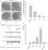
Excess of Gqβ over Gqα is required to prevent production of spontaneous bumps in the dark. (A) Immunogold EM analysis of a cross-section of a single rhabdomere, using a Gqα antibody that was applied to dark adapted wild-type flies and Gβ mutants (bar 500 nm). (B) Number of mean gold particles in cross-sections of 20 different single rhabdomeres. Error bars are SEM. (C) Whole-cell voltage clamp recordings of spontaneous bumps observed in complete darkness of various mutants as indicated. (D) Histogram plotting the mean bump frequency of the various mutants. Error bars are SEM. Note the high spontaneous bump frequency of Gβ hetrozygote compared to the reduced bump frequency of the Gqα/Gβ double hetrozygote mutant (modified from Elia et al., 2005).
Figure 8
Figure 8
Slow response termination composed of bumps characterizes norpA mutants. (A,B) Upper panels: Whole-cell voltage clamp recordings of quantum bumps in response to continues dim light in wild-type and the weak allele of norpA, norpAP57 mutant flies. (A,B) Lower panels: Whole-cell voltage clamp recordings of normalized macroscopic responses of wild-type and the corresponding mutants in response to 200-ms light pulses. In contrast to the fast response termination of wild-type, slow termination of the light response of norpAP57 mutant flies is revealed. This slow response termination can be resolved into continuous production of bumps in the dark at a later time (inset, at higher magnification). (C) Electroretinogram (ERG) responses showing superimposed traces recorded from wild-type and norpAP76 (a weak norpA allele) to a brief flash (red arrow) and continuous light. The graph plots the relative steady state amplitude of the ERG to prolonged lights as a function of relative light intensity. The ERG responses of norpAP76 to a brief flash and to continuous light are indistinguishable.
Figure 9
Figure 9
The phosphoinositide cycle. In the phototransduction cascade, light triggers the activation of phospholipase Cβ (PLCβ). This catalyzes hydrolysis of the membrane phospholipid PIP2 into InsP3 and DAG. DAG is transported by endocytosis to the endoplasmic reticulum and inactivated by phosphorylation converting it into phosphatidic acid (PA) via DAG kinase (DGK) and to CDP-DAG via CDP-DAG syntase. Subsequently, CDP-DAG is converted into phosphatidylinositol (PI), which is transferred back to the microvillar membrane, by the PI transfer protein. PIP and PIP2 are produced at the microvillar membrane by PI kinase and PIP kinase, respectively. There are probably two PIP kinases (PIPK I, PIPK II, which are unified in the scheme). PA can also be converted back to DAG by lipid phosphate phosphohydrolase. PA is also produced from phosphatidylcholine (PC) by phospholipase D (PLD). DAG is also hydrolyzes by DAG lipase into poly unsaturated fatty acids (PUFA).
Figure 10
Figure 10
The electrophysiological properties of WT, trp and trpl mutants. (A) Whole-cell voltage clamp recordings of quantum bumps in response to continuous dim light in wild-type, trpl302 and trpP343 null mutant flies. Highly reduced amplitude of trpP343 bumps is observed. (B) Whole-cell voltage clamp recordings in response to a 3-s light pulse of WT and the corresponding mutants. The transient response of the trpP343 mutant is observed. (C) A family of light-induced currents to 20-ms light pulse at voltage steps of 3 mV measured around Erev. (D) Histogram plotting the mean Erev of WT and the various mutants, error bars are SEM. Erev of wild-type is between the positive Erev of trpl302, which expresses only TRP and the Erev of trpP343 mutant, which expresses only TRPL.
Figure 11
Figure 11
The inaCP209 and inaDP215 mutants reveal slow response termination of the macroscopic response to light and of the single bumps. (A–C) Upper panels: Whole-cell voltage clamp quantum bump responses to continues dim light in wild-type, inaCP209 and inaDP215 mutant flies. A slow termination of the bumps is observed in inaCP209 and inaDP215 mutant flies. (A–C) Lower panels: Whole-cell voltage clamp recordings of normalized responses to a 500-ms light pulse of the above mutants. A slow termination of macroscopic response is observed in inaCP209 null mutant and in the inaDP215 mutant in which the binding of INAD to TRP is disrupted (Chevesich et al., ; Shieh and Zhu, 1996).

Similar articles

Cited by

References

    1. Acharya J. K., Jalink K., Hardy R. W., Hartenstein V., Zuker C. S. (1997). InsP3 receptor is essential for growth and differentiation but not for vision in Drosophila. Neuron 18, 881–88710.1016/S0896-6273(00)80328-1 - DOI - PubMed
    1. Adamski F. M., Zhu M. Y., Bahiraei F., Shieh B. H. (1998). Interaction of eye protein kinase C and INAD in Drosophila. Localization of binding domains and electrophysiological characterization of a loss of association in transgenic flies. J. Biol. Chem. 273, 17713–1771910.1074/jbc.273.28.17713 - DOI - PubMed
    1. Alloway P. G., Dolph P. J. (1999). A role for the light-dependent phosphorylation of visual arrestin. Proc. Natl. Acad. Sci. U. S. A. 96, 6072–607710.1073/pnas.96.11.6072 - DOI - PMC - PubMed
    1. Alloway P. G., Howard L., Dolph P. J. (2000). The formation of stable rhodopsin-arrestin complexes induces apoptosis and photoreceptor cell degeneration. Neuron 28, 129–13810.1016/S0896-6273(00)00091-X - DOI - PubMed
    1. Arnold D. B., Clapham D. E. (1999). Molecular determinants for subcellular localization of PSD-95 with an interacting K+ channel. Neuron 23, 149–15710.1016/S0896-6273(00)80761-8 - DOI - PubMed

LinkOut - more resources