. 2009 Jun 22:2:5.
doi: 10.3389/neuro.02.005.2009. eCollection 2009.

Second and third generation voltage-sensitive fluorescent proteins for monitoring membrane potential

Affiliations

Second and third generation voltage-sensitive fluorescent proteins for monitoring membrane potential

Amelie Perron et al. Front Mol Neurosci. .

Abstract

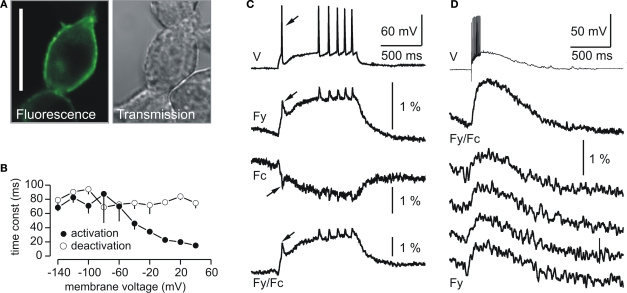
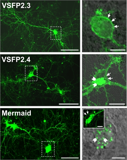
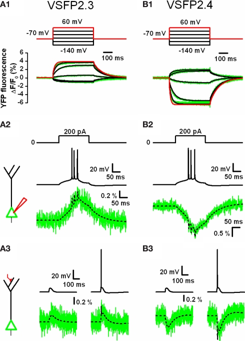
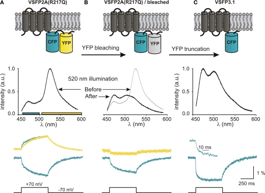
Over the last decade, optical neuroimaging methods have been enriched by engineered biosensors derived from fluorescent protein (FP) reporters fused to protein detectors that convert physiological signals into changes of intrinsic FP fluorescence. These FP-based indicators are genetically encoded, and hence targetable to specific cell populations within networks of heterologous cell types. Among this class of biosensors, the development of optical probes for membrane potential is both highly desirable and challenging. A suitable FP voltage sensor would indeed be a valuable tool for monitoring the activity of thousands of individual neurons simultaneously in a non-invasive manner. Previous prototypic genetically-encoded FP voltage indicators achieved a proof of principle but also highlighted several difficulties such as poor cell surface targeting and slow kinetics. Recently, we developed a new series of FRET-based Voltage-Sensitive Fluorescent Proteins (VSFPs), referred to as VSFP2s, with efficient targeting to the plasma membrane and high responsiveness to membrane potential signaling in excitable cells. In addition to these FRET-based voltage sensors, we also generated a third series of probes consisting of single FPs with response kinetics suitable for the optical imaging of fast neuronal signals. These newly available genetically-encoded reporters for membrane potential will be instrumental for future experimental approaches directed toward the understanding of neuronal network dynamics and information processing in the brain. Here, we review the development and current status of these novel fluorescent probes.

Keywords: fluorescence; fluorescent proteins; genetically-encoded voltage sensors; neuronal circuit dynamics; neurons; optical imaging; patch clamp.


Figures

Figure 1
Figure 1
Plasma membrane expression and fluorescence response properties of VSFP2.1. (A) Confocal fluorescence and transmission images of PC12 cells transfected with VSFP2.1. Note the targeting of VSFP2.1 to the plasma membrane. Scale bar is 25 μm. (B) Voltage dependence of apparent activation and deactivation time constants of VSFP2.1 fluorescence signals in PC12 cells at 35°C. (C,D) VSFP2.1 fluorescence responses to physiological neuronal membrane signals. VSFP2.1-expressing PC12 cells were voltage-clamped with membrane voltage traces recorded from olfactory mitral cells that were stimulated to generate a series of action potentials by direct current injection (C) or by electrical stimulation of the olfactory nerve (D). Traces in (C) and upper trace in (D) are the average of 50 and 90 sweeps, respectively. The lower four traces in (D) are single sweeps. Traces show membrane potential (V), yellow fluorescence (Fy), cyan fluorescence (Fc) and the ratio of yellow and cyan fluorescence (Fy/Fc). Fluorescence signals were digitally low pass filtered (0.2 kHz) and were not corrected for photobleaching. Recordings were done at 35°C (from Dimitrov et al., 2007).
Figure 2
Figure 2
Second generation voltage-sensitive fluorescent proteins. (A) Alignment of the amino acid sequences of VSFP2.1 variants. The C-terminal residues and downstream segment (240–254) of the VSD of Ci-VSP are shown in gray and italic text, respectively. FPs are mCerulean (blue), Citrine/mCitrine (yellow), mKate2 (red), mUKG (green) and mKOκ (orange). (B) Acceptor (upper color traces) and donor (lower color traces) signals of PC12 cells in response to a family of 500 ms voltage steps from a holding potential of −70 mV to test potentials of −140 to +60 mV recorded at 35°C. Traces corresponding to a depolarization to +60 mV are indicated by arrows (from Mutoh et al., 2009).
Figure 3
Figure 3
Expression pattern of VSFP2.1 variants in transfected hippocampal neurons. Primary hippocampal cultures derived from mouse E18 embryos were transfected with either VSFP2.3, VSFP2.4 or Mermaid 6 days after plating and imaged by confocal fluorescence microscopy 1 week later. Fluorescence was allowed to saturate locally to optimize the visualization of neuronal processes. Boxed areas in the left panel are shown magnified on the right panel. Arrows indicate cell surface expression while arrowheads show intracellular expression. Note the targeting of VSFP2.3 and VSFP2.4 to the plasma membrane and Mermaid-associated intracellular aggregates in magnified views. The insert in the lower right image shows an example of a cell with clear expression of Mermaid at the cell surface. Scale bars are 50 and 10 μm for left and right panels, respectively.
Figure 4
Figure 4
Computer simulations of VSFP2.3 and VSFP2.4 optical response signals in cortical layer 5 (L5) pyramidal neurons. (A1) Family of YFP fluorescence (ΔF/F0) responses (lower panel; green traces) to 500 ms voltage steps (upper panel) from a holding membrane voltage of −70 mV to test potentials of −140 to +60 mV recorded from voltage-clamped PC12 cells expressing VSFP2.3. Overlaid to the experimental traces are simulated traces obtained from a VSFP2.3 model wherein the kinetics are represented as an eight state Markov process as given in (Akemann et al., 2009). The simulated response to +60 mV is highlighted in red. (A2) Predicted VSFP2.3 fluorescence signal (ΔF/F0; lower panel) in response to an action potential burst (middle panel) in the cell body of a simulated L5 pyramidal neuron evoked by constant current injection (upper panel). The schematic to the left depicts the neuron with a point current source attached to the cell body (red electrode). The electrical response was simulated using a conductance-based model of a reconstructed rat L5 neuron given by Mainen and Sejnowski (1996). VSFP2.3 was homogenously inserted within the membrane (all 177 compartments) at a density of 800 units/μm2. The fluorescence signal (lower panel) represents the response of VSFP2.3 in the somatic membrane with (green) or without (dashed black) photon quantum noise calculated assuming a sampling rate of 2 kHz and excitation light level that bleaches GFP within 10 s of illumination. (A3) VSFP2.3 fluorescence signal (ΔF/F0; lower panel) as predicted by the simulation in response to a subthreshold (left column) or suprathreshold (right column) activation of a distal synaptic conductance (schematically depicted to the left with the activated synapse in red). The voltage signal in the cell body (upper row left: EPSP; upper row right: EPSP plus evoked action potential) together with the associated fluorescence signals (lower row), with (green) or without (dashed black) photon quantum noise are shown. (B1–B3) Same as in (A1–A3), but using a model of VSFP2.4 instead of VSFP2.3. VSFP2.4 was simulated as an eight state Markov chain model analogously to VSFP2.3. For methodological details see Akemann et al. (2009).
Figure 5
Figure 5
Development of third generation voltage-sensitive fluorescent proteins. The membrane topology of VSFP2A(R217Q) (A), VSFP2A(R217Q) after acceptor photobleaching (B) and VSFP3.1 (C) is illustrated in top panels. Emission spectra recorded for each constructs using 440-nm excitation light are shown below. The lower panel shows the depolarization-induced fluorescence signals recorded in the yellow and cyan channels. For VSFP2A(R217Q), a scaled mirror image of the cyan signal is shown aligned with the yellow signal; note the fast CFP component. For VSFP3.1, the top trace represents the onset of the fluorescence signal at a magnified time scale (from Lundby et al., 2008).
Figure 6
Figure 6
Expression pattern of VSFP3.1 in cultured mouse hippocampal neurons. Primary hippocampal cultures were transfected with VSFP3.1 6 days after plating and confocal images were taken 6 days later. Overviews and magnified views of VSFP3.1 expression in transfected hippocampal neurons are shown. The boxed region in the left panel is shown magnified on the right panel. Arrows indicate plasma membrane expression whereas the arrowhead shows intracellular expression. Scale bar is 50 μm (upper left and lower panel) and 10 μm (upper right).

References

    1. Akemann W., Lundby A., Mutoh H., Knöpfel T. (2009). Effect of voltage sensitive fluorescent proteins on neuronal excitability. Biophys. J. 96, 3959–397610.1016/j.bpj.2009.02.046 - DOI - PMC - PubMed
    1. Ataka K., Pieribone V. A. (2002). A genetically targetable fluorescent probe of channel gating with rapid kinetics. Biophys. J. 82, 509–51610.1016/S0006-3495(02)75415-5 - DOI - PMC - PubMed
    1. Baker B. J., Kosmidis E. K., Vucinic D., Falk C. X., Cohen L. B., Djurisic M., Zecevic D. (2005). Imaging brain activity with voltage- and calcium-sensitive dyes. Cell. Mol. Neurobiol. 25, 245–28210.1007/s10571-005-3059-6 - DOI - PMC - PubMed
    1. Baker B. J., Lee H., Pieribone V. A., Cohen L. B., Isacoff E. Y., Knöpfel T., Kosmidis E. K. (2007). Three fluorescent protein voltage sensors exhibit low plasma membrane expression in mammalian cells. J. Neurosci. Methods 161, 32–3810.1016/j.jneumeth.2006.10.005 - DOI - PubMed
    1. Baker B. J., Mutoh H., Dimitrov D., Akemann W., Perron A., Iwamoto Y., Jin L., Cohen L. B., Isacoff E. Y., Pieribone V. A., Hughes T., Knöpfel T. (2008). Genetically encoded fluorescent sensors of membrane potential. Brain Cell Biol. 36, 53–6710.1007/s11068-008-9026-7 - DOI - PMC - PubMed