

Mobile phone based clinical microscopy for global health applications
- PMID: 19623251
- PMCID: PMC2709430
- DOI: 10.1371/journal.pone.0006320
Mobile phone based clinical microscopy for global health applications
Abstract
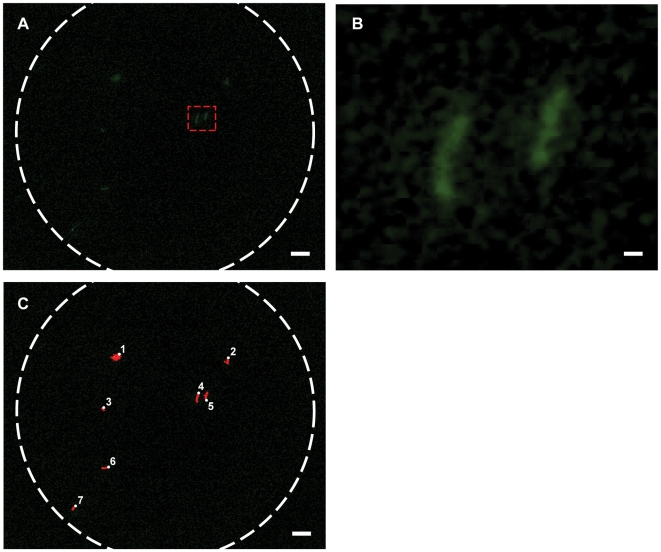
Light microscopy provides a simple, cost-effective, and vital method for the diagnosis and screening of hematologic and infectious diseases. In many regions of the world, however, the required equipment is either unavailable or insufficiently portable, and operators may not possess adequate training to make full use of the images obtained. Counterintuitively, these same regions are often well served by mobile phone networks, suggesting the possibility of leveraging portable, camera-enabled mobile phones for diagnostic imaging and telemedicine. Toward this end we have built a mobile phone-mounted light microscope and demonstrated its potential for clinical use by imaging P. falciparum-infected and sickle red blood cells in brightfield and M. tuberculosis-infected sputum samples in fluorescence with LED excitation. In all cases resolution exceeded that necessary to detect blood cell and microorganism morphology, and with the tuberculosis samples we took further advantage of the digitized images to demonstrate automated bacillus counting via image analysis software. We expect such a telemedicine system for global healthcare via mobile phone -- offering inexpensive brightfield and fluorescence microscopy integrated with automated image analysis -- to provide an important tool for disease diagnosis and screening, particularly in the developing world and rural areas where laboratory facilities are scarce but mobile phone infrastructure is extensive.
Conflict of interest statement
Figures


References
-
- National Electrical Manufacturers Association. Digital Imaging and Communications in Medicine (DICOM) standard. 2008.
-
- Steingart KR, Henry M, Ng V, Hopewell PC, Ramsay A, et al. Fluorescence versus conventional sputum smear microscopy for tuberculosis: a systematic review. Lancet Infect Dis. 2006;6:570–581. - PubMed
-
- World Health Organization. Geneva, Switzerland: 2006. Informal consultation on quality control of malaria microscopy.
-
- World Health Organization- Regional Office for the Western Pacific. Manilla, Philippines: 2003. Quality assurance of sputum microscopy in DOTS programmes.
-
- Chui DHK, Steinberg MH. Laboratory Diagnosis of Hemoglobinopathies and Thalassemias. Hematology: Basic Principles and Practice, 4th ed. 2004. 4th ed:Churchill Livingstone.
Publication types
MeSH terms
LinkOut - more resources
Full Text Sources
Other Literature Sources
Medical
