

doi: 10.1016/j.jsmc.2007.05.010.
The Development of Circadian Rhythms: From Animals To Humans
Affiliations
- PMID: 19623268
- PMCID: PMC2713064
- DOI: 10.1016/j.jsmc.2007.05.010
Item in Clipboard
The Development of Circadian Rhythms: From Animals To Humans
Sleep Med Clin.
.
No abstract available
Figures
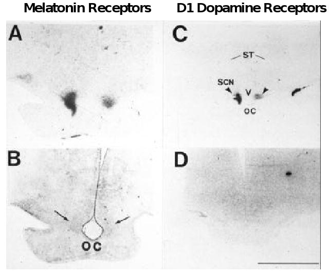
A. Localization of [125I]melatonin binding to the SCN of an 18-week gestation human fetus. Specific labeling is shown in black. B. The stained section used to generate the autoradiograph in A. Reproduced by permission from ref . C. Localization of [125I]SKF38393 binding to D1 dopamine receptors in the SCN of a 20-week post conceptual human infant. Specific labeling is shown in black. D. Non-specific labeling. Reproduced by permission from ref . OC, optic chiasm; ST, striatum. Arrows identify the SCN.
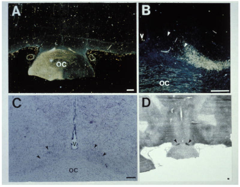
Innervation of the SCN by the retinohypothalamic tract (RHT) in a newborn baboon infant. A. Low-power image showing labeling of retinal fibers in the optic chiasm by horseradish peroxidase. B. Adjacent tissue section showing the location of the SCN. C. High power image showing projections of the RHT into the right SCN. D. Autoradiographic image generated from [14C]2-deoxyglocose uptake studies showing that light exposure at night induces increases in SCN metabolic activity. Areas of increased uptake are dark. Arrows identify the SCN. Scale bar = 5 mm. Reproduced by permission from ref .
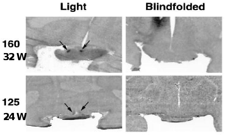
Autoradiograph images of preterm baboon brain sections showing SCN DG uptake after light exposure at night. Animals shown were PC 160 or 125 and were either blindfolded or directly exposed to light. The images are obtained from mid-SCN levels. Arrows identify the SCN image. Reproduced by permission from ref .
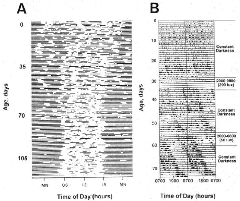
Rest-activity patterns of a newborn human (A) and a baboon (B) infant (right). Double-plotted actograms are shown. In A, dark bars represent sleep. In B, dark bars represent activity. Please note that the circadian phase of the baboons infant was shifted by exposure to a 200 lux reversed light-dark cycle at 30 days of age, and much less so by 50 lux of exposure at 55 days of age. Reproduced by permission from ref .
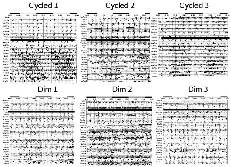
Actograms of rest-activity in representative infants exposed to cycled lighting in the (top panels) or constant dim light (bottom panels). Dark bars represent activity; the same activity scale is used in each plot. The time of day is shown on top. The thick dark line in middle of plots depicts the date of discharge. Note that distinct patterns of rest and activity in the infants are more apparent after discharge in infants exposed to cycled lighting than dim lighting before discharge from the hospital. In infants exposed to dim lighting, day-night differences in rest-activity patterns in synchrony with the light-dark cycle are generally not apparent until about 20 days after discharge from the hospital.
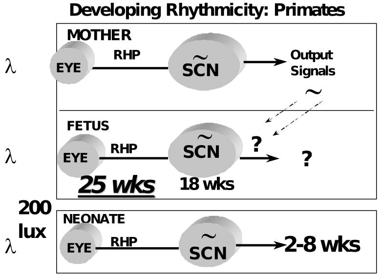
Schematic representation of primate circadian system development based on studies of non-human primates. Estimated human ages are given.
Similar articles
-
Development of circadian rhythms in mammalian systems.Biochem J. 2024 Dec 23;481(24):1967-1976. doi: 10.1042/BCJ20210060. Biochem J. 2024. PMID: 39714414 Review.
-
The role of temperature on the development of circadian rhythms in honey bee workers.PeerJ. 2024 Mar 15;12:e17086. doi: 10.7717/peerj.17086. eCollection 2024. PeerJ. 2024. PMID: 38500530 Free PMC article.
-
Perinatal development of human circadian rhythms.Prog Brain Res. 1996;111:217-26. doi: 10.1016/s0079-6123(08)60410-0. Prog Brain Res. 1996. PMID: 8990917 Review.
-
Ubiquitous light-emitting diodes: Potential threats to retinal circadian rhythms and refractive development.Sci Total Environ. 2023 Mar 1;862:160809. doi: 10.1016/j.scitotenv.2022.160809. Epub 2022 Dec 9. Sci Total Environ. 2023. PMID: 36502986 Review.
-
Importance of Circadian Rhythms in the Ocular Surface.Biomolecules. 2024 Jul 4;14(7):796. doi: 10.3390/biom14070796. Biomolecules. 2024. PMID: 39062510 Free PMC article. Review.
Cited by
-
Transcription and Maturation of mRNA in Dinoflagellates.Microorganisms. 2013 Nov 1;1(1):71-99. doi: 10.3390/microorganisms1010071. Microorganisms. 2013. PMID: 27694765 Free PMC article. Review.
-
Breast Milk and the Importance of Chrononutrition.Front Nutr. 2022 May 12;9:867507. doi: 10.3389/fnut.2022.867507. eCollection 2022. Front Nutr. 2022. PMID: 35634367 Free PMC article. Review.
-
Ultradian rhythms in accelerometric and autonomic data vary based on seizure occurrence in paediatric epilepsy patients.Brain Commun. 2024 Feb 12;6(2):fcae034. doi: 10.1093/braincomms/fcae034. eCollection 2024. Brain Commun. 2024. PMID: 38454964 Free PMC article.
-
Rhythms of life: circadian disruption and brain disorders across the lifespan.Nat Rev Neurosci. 2019 Jan;20(1):49-65. doi: 10.1038/s41583-018-0088-y. Nat Rev Neurosci. 2019. PMID: 30459365 Free PMC article. Review.
-
Birth, love, and fear: Physiological networks from pregnancy to parenthood.Compr Psychoneuroendocrinol. 2022 Apr 26;11:100138. doi: 10.1016/j.cpnec.2022.100138. eCollection 2022 Aug. Compr Psychoneuroendocrinol. 2022. PMID: 35757173 Free PMC article. Review.
References
-
- Panda S, Hogenesch JB, Kay SA. Circadian rhythms from flies to human. Nature. 2002;417(6886):329–35. - PubMed
-
- Ko CH, Takahashi JS. Molecular components of the mammalian circadian clock. Hum Mol Genet. 2006;15(2):R271–7. - PubMed
-
- Maywood ES, O'Neill J, Wong GK, Reddy AB, Hastings MH. Circadian timing in health and disease. Prog Brain Res. 2006;153:253–69. - PubMed
-
- Moore-Ede MC, Czeisler CA, Richardson GS. Circadian timekeeping in health and disease. Part 1. Basic properties of circadian pacemakers. NEnglJMed. 1983;309(8):469–76. - PubMed
-
- Moore-Ede MC, Czeisler CA, Richardson GS. Circadian timekeeping in health and disease. Part 2. Clinical implications of circadian rhythmicity. NEnglJMed. 1983;309(9):530–6. - PubMed
Grants and funding
LinkOut - more resources
Full Text Sources
