. 2008 Oct;105(40):686-92.
doi: 10.3238/arztebl.2008.0686. Epub 2008 Oct 3.

Mastocytosis: a disease of the hematopoietic stem cell

Mastocytosis: a disease of the hematopoietic stem cell

Hans-Peter Horny et al. Dtsch Arztebl Int. 2008 Oct.

Abstract

Introduction: Mastocytosis is an unusual clonal disease of the hematopoietic stem cell.

Methods: This article is based on a selective literature search and on the authors' clinical and pathological experience.

Results: The clinical manifestations of mastocytosis range from cutaneous mastocytosis, a common, prognostically favorable presentation, to mast cell leukemia, a rare, life-threatening disease. The mediator-induced symptoms usually respond well to H1 antihistamines. Therapeutic standards for cytoreduction in the progressive, systemic forms of mastocytosis are still lacking.

Discussion: Because some of the manifestations of mastocytosis are nonspecific and can be mimicked by other diseases, there is a risk of two types of diagnostic error: Mastocytosis may remain undiagnosed when it is actually present, or it may be diagnosed even though morphological and molecular findings rule out mastocytosis. Well-defined criteria should be used to differentiate mastocytosis from other diseases with a similar clinical presentation.

Keywords: anaphylactic reaction; diagnosis; differential diagnosis; mastocytosis; urticaria.


Figures

Figure 1
Figure 1
Typical maculopapular skin lesions (urticaria pigmentosa) in adults with cutaneous or systemic mastocytosis
Figure 2
Figure 2
The maculopapular skin lesions are usually larger in children than in adults; as a rule there is no systemic involvement
Figure 3
Figure 3
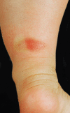
Solitary mastocytoma of the distal lower leg. Mastocytomas are brown or brownish-red, usually well defined and raised. After mechanical irritation with a wooden spatula, the various mastocytic skin lesions show reddening and urticarial swelling (Darier’s sign). After the reddening has worn off it may be followed by a white anemic discoloration of a few minutes in duration, as can be seen here to the left of the mastocytoma
Figure 4
Figure 4
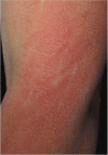
In diffuse cutaneous mastocytosis the skin is uniformly yellowish-red in color and thickened; histological examination shows marked mast cell proliferation in the dermis
Figure 5
Figure 5
Focal infiltration of the bone marrow by systemic mastocytosis. The mast cells are densely packed, mostly spindle-shaped, and hypogranulated. This meets the major criterion (compact mast cell infiltrate) and one of the minor criteria (spindle-shaped mast cells) of the WHO for the diagnosis of systemic mastocytosis
Figure 6
Figure 6
Mild diffuse proliferation of partially spindle-shaped, often hypogranulated tissue mast cells without the formation of compact (diagnostic) infiltrates. This pattern is typical for adult patients with the skin lesions of cutaneous mastocytosis (urticaria pigmentosa) in whom the bone marrow is investigated for staging purposes. Clarification by means of special immunohistochemical stains (particularly CD25) and molecular-genetic investigation for confirmation/exclusion of a Kit mutation in exon 17 is necessary in such cases. If an atypical phenotype with mast cell expression of CD25 is demonstrated and a mutation is found, then together with the spindle-shaped cells three minor diagnostic criteria are fulfilled and the diagnosis of indolent systemic mastocytosis with involvement of skin and bone marrow is established
Figure 7
Figure 7
Markedly hypercellular bone marrow with diffuse compact infiltrates of a lymphocytic lymphoma; predominance of small lymphatic cells and subtotal depletion of fat cells and normal blood cell precursors. Note the diffuse proliferation of medium-sized, round, always loosely scattered tissue mast cells with numerous metachromatic granules. Typical appearance of reactive mast cell proliferation (mast cell hyperplasia), clearly distinct in this example from the findings in systemic mastocytosis

Comment in

References

    1. Ehrlich P. Beiträge zur Kenntniss der granulierten Bindegewebszellen und der eosinophilen Leukocythen. Arch Anat Physiol. 1879;3:166–169.
    1. Galli SJ, Tsai M. Mast cells: versatile regulators of inflammation, tissue remodeling, host defense and homeostasis. J Dermatol Sci. 2008;49:7–19. - PMC - PubMed
    1. Hartmann K, Henz BM. Mastocytosis: recent advances in defining the disease. Br J Dermatol. 2001;144:682–695. - PubMed
    1. Valent P, Horny H-P, Li CY, et al. World Health Organization (WHO) classification of tumors. Mastocytosis (mast cell disease) In: Jaffe ES, Harris NL, Stein H, Vardiman JW, editors. Pathology and genetics of tumors of haematopoietic and lymphoid tissues. Lyon: IARC Press; 2001. pp. 291–302.
    1. Valent P, Horny H-P, Escribano L, Longley JB, Li CY, Schwartz LB, et al. Diagnostic criteria and classification of mastocytosis: a consensus proposal. Conference report of „Year 2000 Working Conference on Mastocytosis“. Leuk Res. 2001;25:603–625. - PubMed