Review
. 2009 Sep;238(9):2139-48.
doi: 10.1002/dvdy.22034.

Signaling in the third dimension: the peripodial epithelium in eye disc development

Affiliations
Review

Signaling in the third dimension: the peripodial epithelium in eye disc development

Mardelle Atkins et al. Dev Dyn. 2009 Sep.

Abstract

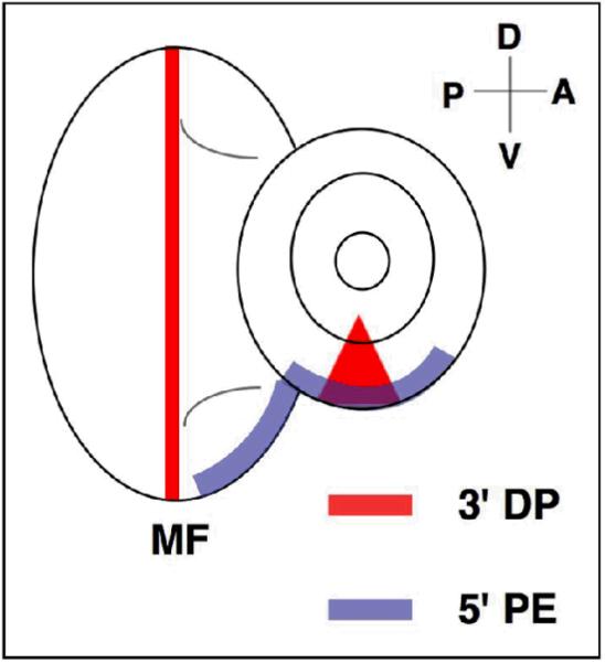
The eye-antennal imaginal disc of Drosophila melanogaster has often been described as an epithelial monolayer with complex signaling events playing out in two dimensions. However, the imaginal disc actually comprises two opposing epithelia (the peripodial epithelium, or PE, and the disc proper, or DP) separated by a lumen to form a sac-like structure. Recent studies expose complex molecular interactions between the PE and the DP, and reveal dynamic communication between the two tissues. Further findings suggest the PE makes important contributions to DP development by acting as a source of signaling molecules as well as cells. Here we summarize those findings and highlight implications for further research.


Figures

Figure 1
Figure 1. The eye-antennal imaginal disc is a dynamic structure in Drosophila development
The central black line depicts the passage of time in hours. Drosophila development is schematized to show the relative growth of the whole animal (above time line) and the eye-antennal disc (below time line). Key events for each stage are detailed in text (bottom). Embryonic development (red) takes place over 24 hours. During this time the eye-antennal imaginal disc precursors are specified. The eye-antennal disc is recoverable as a distinct structure during first instar (orange). At this stage the antennal disc is not distinct from the eye. During second instar (green), the disc proliferates, the D/V axis is specified, and the antennal disc becomes distinct. At the onset of third instar (blue) the MF initiates at the posterior margin (indigo line). The disc continues to grow and differentiation occurs progressively throughout the third instar. By late third instar the MF has crossed most of the eye field, and more posterior ommatidia are undergoing rotation. During pupation (violet) the antennal disc (not depicted) separates from the eye disc and both discs evert and fuse with head cuticle tissue and the labral and clypeolabral discs to produce the adult head. The final steps of differentiation are completed during pupation.
Figure 2
Figure 2. The squamous PE overlies the columnar DP in the eye imaginal disc
A) The eye-antennal imaginal disc is depicted at the level of the DP. A/P and D/V axes are indicated in the upper right corner. The eye disc proper (pink), the antennal disc proper (orange), and the optic stalk (OS) are illustrated. X and Y represent cross sections through the disc corresponding to the dashed lines. B) Sections of the disc corresponding to X and Y in panel A are depicted. The thin squamous PE (green) is shown overlying the DP (pink or orange), separated by a lumen. MF indicates the apical constriction corresponding to the morphogenetic furrow. The cuboidal margin cells are shown in blue (blue arrow). C-E) Mouse anti-Discs large staining (1:500, Developmental Studies Hybridoma Bank) labels septate junctions, revealing the apical cell membrane profile of eye-antennal imaginal disc cells. C) A thin optical section taken using confocal microscopy at the level of the PE reveals the large squamous cell profile. D) A thin optical section of the apical DP reveals the small profiles of columnar cells. The yellow asterisks in C and D mark the region of the disc magnified in E and F to facilitate comparison of the relative sizes of PE and DP cells.
Figure 3
Figure 3. A model linking disc growth and D/V boundary establishment to initiation of the MF
The DP (pink), the PE (green), and the cuboidal margin cells (blue) are shown. In all panels dorsal (D) and ventral (V) are indicated, and posterior is to the left in B-D. Panel A is the cross section indicated by the dashed line in B. A) A second instar disc is represented in cross section to reveal the PE and DP. During second instar Dpp, Hh, and Wg are expressed asymmetrically within the PE (not shown) and their activity refines Dl and Ser expression in the DP. The boundary between Dl and Ser localization establishes activation of Notch at the midline and establishes the D/V boundary in the disc (dark pink). B) Activated Notch at the midline induces Upd expression in the posterior cuboidal margin cells. C) Upd secretion from the posterior margin stimulates DP growth and represses wg expression, particularly in the PE. Loss of Wg at the posterior margin results in expression of odd and drm. D) Drm and Odd inhibit Lines activity in posterior margin cells, de-repressing Bowl, which in turn activates the transcription of hh, initiating the MF.
Figure 4
Figure 4. Ommatidia of the adult eye are arranged precisely and form a line of mirror symmetry about the D/V midline
A) A section through an adult eye reveals the highly ordered array of photoreceptors and ommatidia, and includes the D/V midline (red line). B) The diagram depicts the mirror image chirality that is a result of the integration of planar polarity cues. The solid red line represents the D/V midline or equator.
Figure 5
Figure 5. Anatomy of the adult head of Drosophila melanogaster
The anatomy of the fly head is depicted here for reference. Reporter analysis suggests that the cuticle beneath the eye, the second antennal segment, the vibrissae, maxillary palps, and the frons (names highlighted in green) contain cells that are derived from the PE. The clypeus is derived from the clypeolabral disc, and the labrellum is derived from the labial disc (names in gray).
Figure 6
Figure 6. 5' and 3' dpp enhancers have distinct expression patterns
The 3' DP enhancer expression pattern is shown in red and the 5' PE enhancer is shown in blue. The expression of the 3' enhancer in the eye disc corresponds to the moving wave of differentiation, the morphogenetic furrow (MF).

Similar articles

Cited by

References

    1. Baker NE. Transcription of the segment-polarity gene wingless in the imaginal discs of Drosophila, and the phenotype of a pupal-lethal wg mutation. Development. 1988;102:489–497. - PubMed
    1. Bessa J, Casares F. Restricted teashirt expression confers eye-specific responsiveness to Dpp and Wg signals during eye specification in Drosophila. Development. 2005;132:5011–5020. - PubMed
    1. Bodentstein D. The Postembryonic development of Drosophila melanogaster. In: Demerec M, editor. Biology of Drosophila. John Wiley and Sons, Inc.; New York: 1950. pp. 275–367.
    1. Bras-Pereira C, Bessa J, Casares F. Odd-skipped genes specify the signaling center that triggers retinogenesis in Drosophila. Development. 2006;133:4145–4149. - PubMed
    1. Cavodeassi F, Diez Del Corral R, Campuzano S, Dominguez M. Compartments and organising boundaries in the Drosophila eye: the role of the homeodomain Iroquois proteins. Development. 1999;126:4933–4942. - PubMed

Publication types

LinkOut - more resources