. 2009 Nov 1;125(9):2219-28.
doi: 10.1002/ijc.24611.

Multiple pathways in the FGF signaling network are frequently deregulated by gene amplification in oral dysplasias

Affiliations

Multiple pathways in the FGF signaling network are frequently deregulated by gene amplification in oral dysplasias

Ivy F L Tsui et al. Int J Cancer. .

Abstract

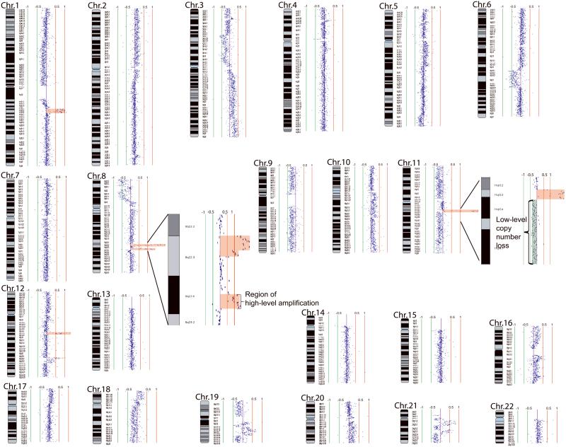
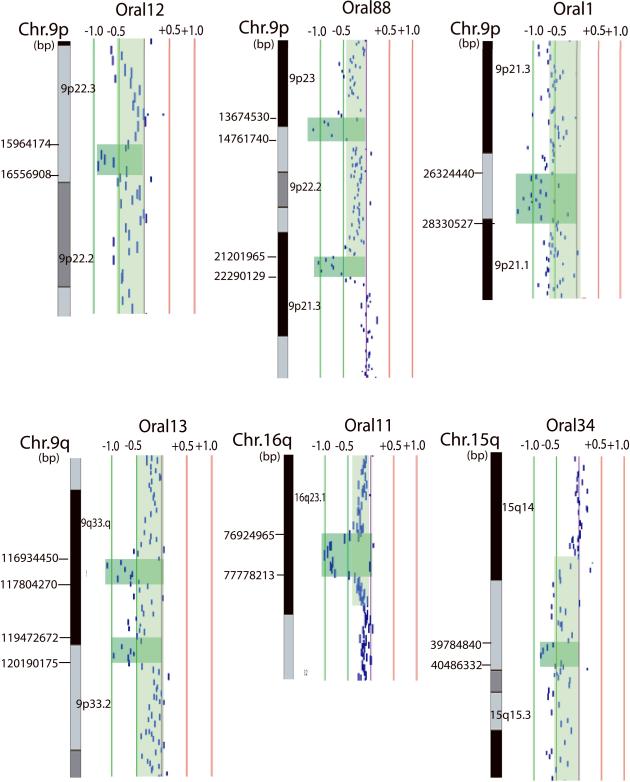
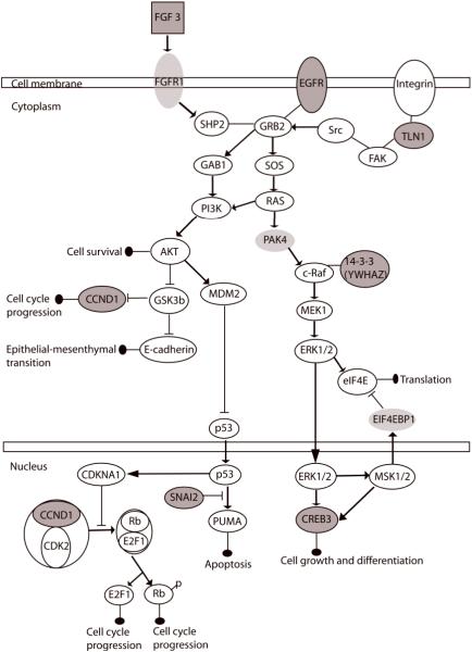
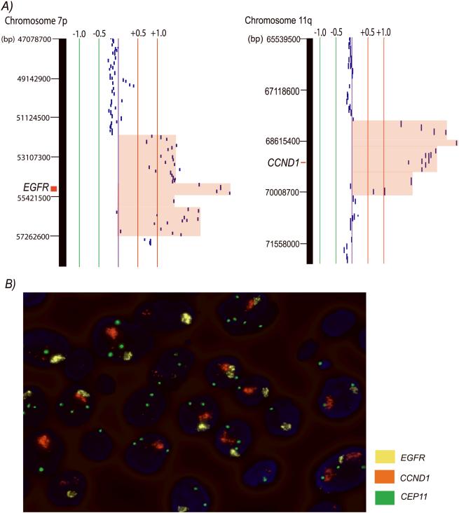
Genetic alteration in oral premalignant lesions (OPLs), the precursors of oral squamous cell carcinomas (OSCCs), may represent key changes in disease initiation and development. We ask if DNA amplification occurs at this early stage of cancer development and which oncogenic pathways are disrupted in OPLs. Here, we evaluated 50 high-grade dysplasias and low-grade dysplasias that later progressed to cancer for gene dosage aberrations using tiling-path DNA microarrays. Early occurrences of DNA amplification and homozygous deletion were frequently detected, with 40% (20/50) of these early lesions exhibiting such features. Expression for 88 genes in 7 recurrent amplicons were evaluated in 5 independent head and neck cancer datasets, with 40 candidates found to be overexpressed relative to normal tissues. These genes were significantly enriched in the canonical ERK/MAPK, FGF, p53, PTEN and PI3K/AKT signaling pathways (p = 8.95 x 10(-3) to 3.18 x 10(-2)). These identified pathways share interactions in one signaling network, and amplification-mediated deregulation of this network was found in 30.0% of these preinvasive lesions. No such alterations were found in 14 low-grade dysplasias that did not progress, whereas 43.5% (10/23) of OSCCs were found to have altered genes within the pathways with DNA amplification. Multitarget FISH showed that amplification of EGFR and CCND1 can coexist in single cells of an oral dysplasia, suggesting the dependence on multiple oncogenes for OPL progression. Taken together, these findings identify a critical biological network that is frequently disrupted in high-risk OPLs, with different specific genes disrupted in different individuals.


Figures

Figure 1
Figure 1
Whole genome tiling-path array profile of a carcinoma in situ (CIS) Oral42. Each data point represents one BAC-derived segment on the array. The log2 signal intensity ratios of a competitive hybridization with pooled male genomic DNA are plotted by SeeGH software. The red and green bar lines are positive and negative log2 signal intensity ratio lines scaled by an increment of 0.5. Data points to the left and right of the centre line represent DNA copy number losses and gains, respectively. Specifically, seven regions of gene amplification on 1q23.2-q23.3, 1q23.3-q24.1, 8q22.2-q22.3, 8q23.1, 11q13.3-q13.4, 12q14.3, and 12q23.2-q23.3 are shaded red. Magnified views of the two amplicons on chromosome 8, and a region of low-level copy number loss and a region of gene amplification on chromosome 11 are shown in two insets.
Figure 2
Figure 2
Graphical representations of region of homozygous deletion in oral lesions. Each data point represents one BAC-derived segment on the array. The detected region of homozygous deletion in each sample is shaded in dark green, whereas single copy loss is shaded in pale green on the corresponding data points. The red and green lines are positive and negative ratio lines scaled by an increment of log2 signal ratios of 0.5.
Figure 3
Figure 3
Multiple disruptions in a single network driven by the mechanism of gene amplification. The top significantly deregulated canonical pathways in oral dysplasias are the ERK/MAPK, FGF, p53, PTEN, and PI3K/AKT signaling pathways, which share common nodes and interplay as a single network. Genes colored in grey with outlined circle were recurrently deregulated by gene amplification in oral dysplasias and significantly overexpressed in independent head and neck cancer datasets, whereas those colored in light grey were found to be amplified only in one preinvasive lesion. Altogether, 25 oral lesions (one progressing low-grade lesions, 14 high-grade dysplasias, and ten OSCCs) exhibited high-level gene amplification of different genes inside this network, contributing to a disruption of 34.2% of all the progressing low-grade dysplasias, high-grade dysplasias, and OSCCs (N = 73).
Figure 4
Figure 4
Co-amplification of EGFR and CCND1 in high-grade dysplasia Oral22. A) The left SeeGH profile represents amplification at 7p11.2 (EGFR locus), whereas the right profile represents amplification at 11q13.3 (CCND1 locus) of sample Oral22. The red and green lines are positive and negative ratio lines scaled by an increment of log2 signal ratios of 0.5. Amplified region was shaded in red. B) FISH visualization of co-amplification of EGFR and CCND1 in single dysplastic cells of Oral22. Sequential FISH was performed with probes mapping to CCND1, EGFR, and the centromeric region of chromosome 11, respectively displayed as orange, yellow, and green signal (original magnification, 1000x). Note the large clusters of amplification signals of CCND1 and EGFR relative to the centromeric region of chromosomes 11 in the same nucleus.

References

    1. Cancer Facts & Figures 2007. American Cancer Society; Atlanta: 2007.
    1. Epstein JB, Gorsky M, Cabay RJ, Day T, Gonsalves W. Screening for and diagnosis of oral premalignant lesions and oropharyngeal squamous cell carcinoma: role of primary care physicians. Can Fam Physician. 2008;54:870–5. - PMC - PubMed
    1. Poh CF, Ng S, Berean KW, Williams PM, Rosin MP, Zhang L. Biopsy and histopathologic diagnosis of oral premalignant and malignant lesions. J Can Dent Assoc. 2008;74:283–8. - PubMed
    1. Warnakulasuriya S, Reibel J, Bouquot J, Dabelsteen E. Oral epithelial dysplasia classification systems: predictive value, utility, weaknesses and scope for improvement. J Oral Pathol Med. 2008;37:127–33. - PubMed
    1. Definition of leukoplakia and related lesions: An aid to studies on oral precancer. Oral Surgery, Oral Medicine, Oral Pathology. 1978;46:518–39. - PubMed

Publication types

MeSH terms