

Detection of basal cell carcinoma using color and histogram measures of semitranslucent areas
- PMID: 19624424
- PMCID: PMC3154079
- DOI: 10.1111/j.1600-0846.2009.00354.x
Detection of basal cell carcinoma using color and histogram measures of semitranslucent areas
Abstract
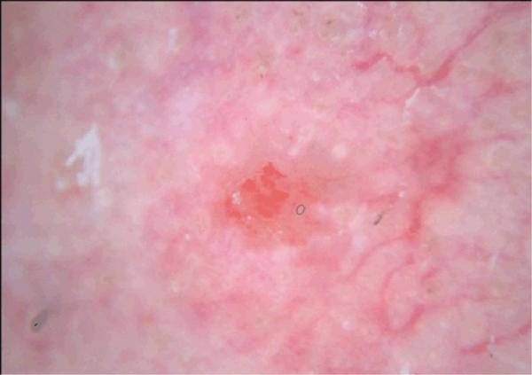
Background: Semitranslucency, defined as a smooth, jelly-like area with varied, near-skin-tone color, can indicate a diagnosis of basal cell carcinoma (BCC) with high specificity. This study sought to analyze potential areas of semitranslucency with histogram-derived texture and color measures to discriminate BCC from non-semitranslucent areas in non-BCC skin lesions.
Methods: For 210 dermoscopy images, the areas of semitranslucency in 42 BCCs and comparable areas of smoothness and color in 168 non-BCCs were selected manually. Six color measures and six texture measures were applied to the semitranslucent areas of the BCC and the comparable areas in the non-BCC images.
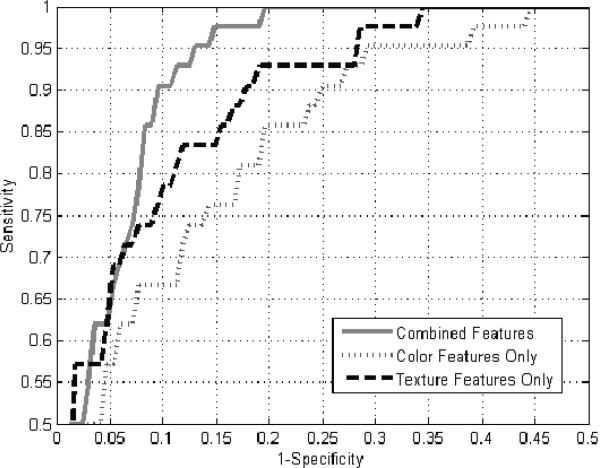
Results: Receiver operating characteristic (ROC) curve analysis showed that the texture measures alone provided greater separation of BCC from non-BCC than the color measures alone. Statistical analysis showed that the four most important measures of semitranslucency are three histogram measures: contrast, smoothness, and entropy, and one color measure: blue chromaticity. Smoothness is the single most important measure. The combined 12 measures achieved a diagnostic accuracy of 95.05% based on area under the ROC curve.
Conclusion: Texture and color analysis measures, especially smoothness, may afford automatic detection of BCC images with semitranslucency.
Figures
Similar articles
-
Detection of granularity in dermoscopy images of malignant melanoma using color and texture features.Comput Med Imaging Graph. 2011 Mar;35(2):144-7. doi: 10.1016/j.compmedimag.2010.09.005. Epub 2010 Oct 30. Comput Med Imaging Graph. 2011. PMID: 21036538 Free PMC article.
-
Visual inspection and dermoscopy, alone or in combination, for diagnosing keratinocyte skin cancers in adults.Cochrane Database Syst Rev. 2018 Dec 4;12(12):CD011901. doi: 10.1002/14651858.CD011901.pub2. Cochrane Database Syst Rev. 2018. PMID: 30521688 Free PMC article.
-
Adaptable texture-based segmentation by variance and intensity for automatic detection of semitranslucent and pink blush areas in basal cell carcinoma.Skin Res Technol. 2016 Nov;22(4):412-422. doi: 10.1111/srt.12281. Epub 2016 Mar 16. Skin Res Technol. 2016. PMID: 26991418
-
Region growing by sector analysis for detection of blue-gray ovoids in basal cell carcinoma.Skin Res Technol. 2013 Aug;19(3):258-64. doi: 10.1111/srt.12036. Epub 2013 Jun 1. Skin Res Technol. 2013. PMID: 23724851 Free PMC article.
-
Exfoliative cytology for diagnosing basal cell carcinoma and other skin cancers in adults.Cochrane Database Syst Rev. 2018 Dec 4;12(12):CD013187. doi: 10.1002/14651858.CD013187. Cochrane Database Syst Rev. 2018. PMID: 30521689 Free PMC article.
Cited by
-
Automated detection of nonmelanoma skin cancer using digital images: a systematic review.BMC Med Imaging. 2019 Feb 28;19(1):21. doi: 10.1186/s12880-019-0307-7. BMC Med Imaging. 2019. PMID: 30819133 Free PMC article.
-
Detection of granularity in dermoscopy images of malignant melanoma using color and texture features.Comput Med Imaging Graph. 2011 Mar;35(2):144-7. doi: 10.1016/j.compmedimag.2010.09.005. Epub 2010 Oct 30. Comput Med Imaging Graph. 2011. PMID: 21036538 Free PMC article.
-
Discrimination of basal cell carcinoma from benign lesions based on extraction of ulcer features in polarized-light dermoscopy images.Skin Res Technol. 2012 Nov;18(4):471-5. doi: 10.1111/j.1600-0846.2011.00595.x. Epub 2012 Feb 22. Skin Res Technol. 2012. PMID: 22356565 Free PMC article.
-
Visual inspection and dermoscopy, alone or in combination, for diagnosing keratinocyte skin cancers in adults.Cochrane Database Syst Rev. 2018 Dec 4;12(12):CD011901. doi: 10.1002/14651858.CD011901.pub2. Cochrane Database Syst Rev. 2018. PMID: 30521688 Free PMC article.
-
Dermoscopy, with and without visual inspection, for diagnosing melanoma in adults.Cochrane Database Syst Rev. 2018 Dec 4;12(12):CD011902. doi: 10.1002/14651858.CD011902.pub2. Cochrane Database Syst Rev. 2018. PMID: 30521682 Free PMC article.
References
-
- Argenziano G, Soyer HP, Chimenti S, Talamini R, Corona R, Sera F, et al. Dermoscopy of pigmented skin lesions: Results of a consensus meeting via the Internet. J Am Acad Dermatol. 2003;48:679–93. - PubMed
-
- Menzies SW, Zalaudek I. Why perform Dermoscopy? The evidence for its role in the routine management of pigmented skin lesions. Arch Dermatol. 2006;142:1211–1222. - PubMed
-
- Vanker AD, Stoecker WV. An expert diagnostic program for dermatology. Comput Biomed Res. 1984;17:241–7. - PubMed
-
- Gonzalez RC, Woods RE. Digital Image Processing. 3rd Ed. Pearson Prentice Hall; Upper Saddle River, NJ: 2008.
Publication types
MeSH terms
Grants and funding
LinkOut - more resources
Full Text Sources
Medical
