

Approximate lesion localization in dermoscopy images
- PMID: 19624428
- PMCID: PMC3152314
- DOI: 10.1111/j.1600-0846.2009.00357.x
Approximate lesion localization in dermoscopy images
Abstract
Background: Dermoscopy is one of the major imaging modalities used in the diagnosis of melanoma and other pigmented skin lesions. Because of the difficulty and subjectivity of human interpretation, automated analysis of dermoscopy images has become an important research area. Border detection is often the first step in this analysis.
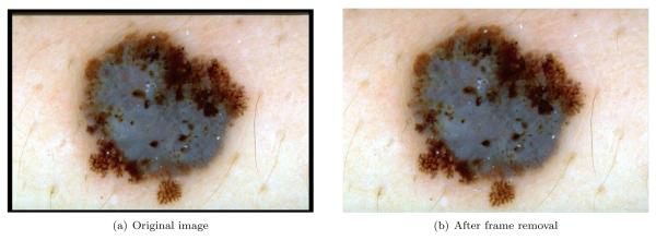
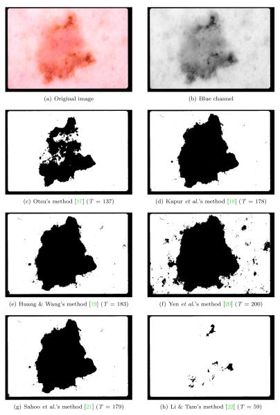
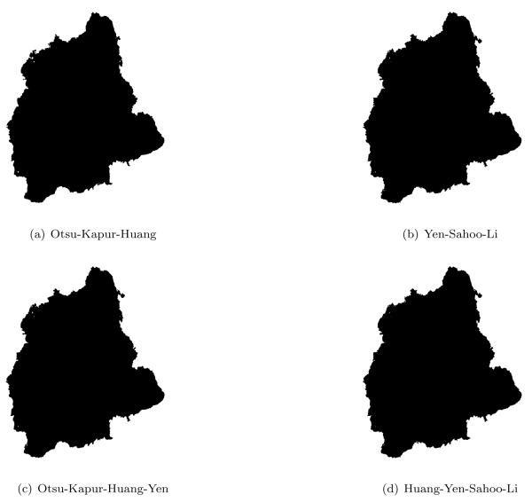
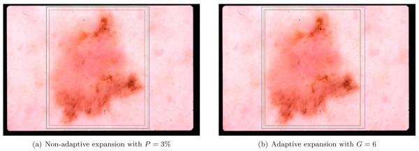
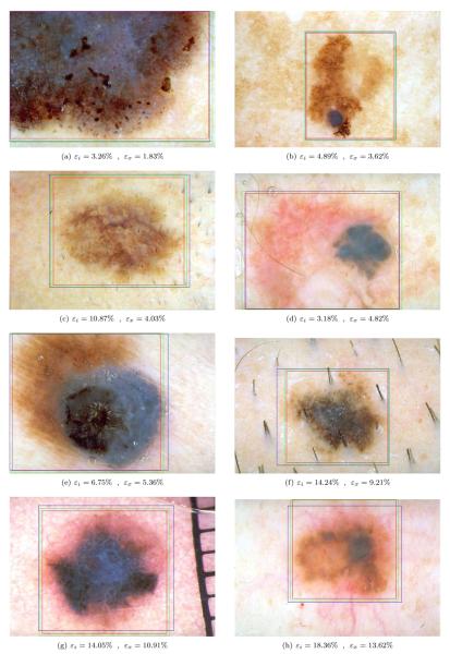
Methods: In this article, we present an approximate lesion localization method that serves as a preprocessing step for detecting borders in dermoscopy images. In this method, first the black frame around the image is removed using an iterative algorithm. The approximate location of the lesion is then determined using an ensemble of thresholding algorithms.
Results: The method is tested on a set of 428 dermoscopy images. The localization error is quantified by a metric that uses dermatologist-determined borders as the ground truth.
Conclusion: The results demonstrate that the method presented here achieves both fast and accurate localization of lesions in dermoscopy images.
Figures
Similar articles
-
Objective evaluation of methods for border detection in dermoscopy images.Annu Int Conf IEEE Eng Med Biol Soc. 2008;2008:3056-9. doi: 10.1109/IEMBS.2008.4649848. Annu Int Conf IEEE Eng Med Biol Soc. 2008. PMID: 19163351
-
Border detection in dermoscopy images using statistical region merging.Skin Res Technol. 2008 Aug;14(3):347-53. doi: 10.1111/j.1600-0846.2008.00301.x. Skin Res Technol. 2008. PMID: 19159382 Free PMC article.
-
Fast density-based lesion detection in dermoscopy images.Comput Med Imaging Graph. 2011 Mar;35(2):128-36. doi: 10.1016/j.compmedimag.2010.07.007. Epub 2010 Sep 17. Comput Med Imaging Graph. 2011. PMID: 20800995
-
Lesion border detection in dermoscopy images using ensembles of thresholding methods.Skin Res Technol. 2013 Feb;19(1):e252-8. doi: 10.1111/j.1600-0846.2012.00636.x. Epub 2012 Jun 7. Skin Res Technol. 2013. PMID: 22676490
-
Lesion border detection in dermoscopy images.Comput Med Imaging Graph. 2009 Mar;33(2):148-53. doi: 10.1016/j.compmedimag.2008.11.002. Epub 2009 Jan 3. Comput Med Imaging Graph. 2009. PMID: 19121917 Free PMC article. Review.
Cited by
-
Analysis of density based and fuzzy c-means clustering methods on lesion border extraction in dermoscopy images.BMC Bioinformatics. 2010 Oct 7;11 Suppl 6(Suppl 6):S26. doi: 10.1186/1471-2105-11-S6-S26. BMC Bioinformatics. 2010. PMID: 20946610 Free PMC article.
References
-
- Jemal A, Siegel R, Ward E, et al. Cancer Statistics, 2008. CA: A Cancer Journal for Clinicians. 2008;58(2):71–96. 2008. - PubMed
-
- Argenziano G, Soyer HP, De Giorgi V, et al. Dermoscopy: A Tutorial. EDRA Medical Publishing & New Media; Milan, Italy: 2002.
-
- Steiner K, Binder M, Schemper M, et al. Statistical Evaluation of Epiluminescence Dermoscopy Criteria for Melanocytic Pigmented Lesions. Journal of American Academy of Dermatology. 1993;29(4):581–588. - PubMed
-
- Binder M, Schwarz M, Winkler A, et al. Epiluminescence Microscopy. A Useful Tool for the Diagnosis of Pigmented Skin Lesions for Formally Trained Dermatologists. Archives of Dermatology. 1995;131(3):286–291. - PubMed
-
- Fleming MG, Steger C, Zhang J, et al. Techniques for a Structural Analysis of Dermatoscopic Imagery. Computerized Medical Imaging and Graphics. 1998;22(5):375–389. - PubMed
Publication types
MeSH terms
Grants and funding
LinkOut - more resources
Full Text Sources
Other Literature Sources
Medical
