Type I interferon receptor controls B-cell expression of nucleic acid-sensing Toll-like receptors and autoantibody production in a murine model of lupus
- PMID: 19624844
- PMCID: PMC2745794
- DOI: 10.1186/ar2771
Type I interferon receptor controls B-cell expression of nucleic acid-sensing Toll-like receptors and autoantibody production in a murine model of lupus
Abstract
Introduction: Systemic lupus erythematosus (SLE) is a chronic autoimmune disease characterized by the production of high-titer IgG autoantibodies directed against nuclear autoantigens. Type I interferon (IFN-I) has been shown to play a pathogenic role in this disease. In the current study, we characterized the role of the IFNAR2 chain of the type I IFN (IFN-I) receptor in the targeting of nucleic acid-associated autoantigens and in B-cell expression of the nucleic acid-sensing Toll-like receptors (TLRs), TLR7 and TLR9, in the pristane model of lupus.
Methods: Wild-type (WT) and IFNAR2-/- mice were treated with pristane and monitored for proteinuria on a monthly basis. Autoantibody production was determined by autoantigen microarrays and confirmed using enzyme-linked immunosorbent assay (ELISA) and immunoprecipitation. Serum immunoglobulin isotype levels, as well as B-cell cytokine production in vitro, were quantified by ELISA. B-cell proliferation was measured by thymidine incorporation assay.
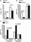
Results: Autoantigen microarray profiling revealed that pristane-treated IFNAR2-/- mice lacked autoantibodies directed against components of the RNA-associated autoantigen complexes Smith antigen/ribonucleoprotein (Sm/RNP) and ribosomal phosphoprotein P0 (RiboP). The level of IgG anti-single-stranded DNA and anti-histone autoantibodies in pristane-treated IFNAR2-/- mice was decreased compared to pristane-treated WT mice. TLR7 expression and activation by a TLR7 agonist were dramatically reduced in B cells from IFNAR2-/- mice. IFNAR2-/- B cells failed to upregulate TLR7 as well as TLR9 expression in response to IFN-I, and effector responses to TLR7 and TLR9 agonists were significantly decreased as compared to B cells from WT mice following treatment with IFN-alpha.
Conclusions: Our studies provide a critical link between the IFN-I pathway and the regulation of TLR-specific B-cell responses in a murine model of SLE.
Figures


Similar articles
-
Maintenance of autoantibody production in pristane-induced murine lupus.Arthritis Res Ther. 2015 Dec 30;17:384. doi: 10.1186/s13075-015-0886-9. Arthritis Res Ther. 2015. PMID: 26717913 Free PMC article.
-
Distinct autoantibody profiles in systemic lupus erythematosus patients are selectively associated with TLR7 and TLR9 upregulation.J Clin Immunol. 2013 Jul;33(5):954-64. doi: 10.1007/s10875-013-9887-0. Epub 2013 Apr 7. J Clin Immunol. 2013. PMID: 23564191
-
DNA-like class R inhibitory oligonucleotides (INH-ODNs) preferentially block autoantigen-induced B-cell and dendritic cell activation in vitro and autoantibody production in lupus-prone MRL-Fas(lpr/lpr) mice in vivo.Arthritis Res Ther. 2009;11(3):R79. doi: 10.1186/ar2710. Epub 2009 May 28. Arthritis Res Ther. 2009. PMID: 19476613 Free PMC article.
-
Toll-like receptor signalling in B cells during systemic lupus erythematosus.Nat Rev Rheumatol. 2021 Feb;17(2):98-108. doi: 10.1038/s41584-020-00544-4. Epub 2020 Dec 18. Nat Rev Rheumatol. 2021. PMID: 33339987 Free PMC article. Review.
-
Emerging roles of TLR7 and TLR9 in murine SLE.J Autoimmun. 2009 Nov-Dec;33(3-4):231-8. doi: 10.1016/j.jaut.2009.10.001. Epub 2009 Oct 21. J Autoimmun. 2009. PMID: 19846276 Review.
Cited by
-
Involvement of TLR7 MyD88-dependent signaling pathway in the pathogenesis of adult-onset Still's disease.Arthritis Res Ther. 2013 Mar 4;15(2):R39. doi: 10.1186/ar4193. Arthritis Res Ther. 2013. PMID: 23497717 Free PMC article.
-
High Interferon Signature Leads to Increased STAT1/3/5 Phosphorylation in PBMCs From SLE Patients by Single Cell Mass Cytometry.Front Immunol. 2022 Jan 28;13:833636. doi: 10.3389/fimmu.2022.833636. eCollection 2022. Front Immunol. 2022. PMID: 35185925 Free PMC article.
-
Meta-analysis of microarray data using a pathway-based approach identifies a 37-gene expression signature for systemic lupus erythematosus in human peripheral blood mononuclear cells.BMC Med. 2011 May 30;9:65. doi: 10.1186/1741-7015-9-65. BMC Med. 2011. PMID: 21624134 Free PMC article.
-
Pristane-Accelerated Autoimmune Disease in (SWR X NZB) F1 Mice Leads to Prominent Tubulointerstitial Inflammation and Human Lupus Nephritis-Like Fibrosis.PLoS One. 2016 Oct 19;11(10):e0164423. doi: 10.1371/journal.pone.0164423. eCollection 2016. PLoS One. 2016. PMID: 27760209 Free PMC article.
-
Plasmacytoid dendritic cells promote rotavirus-induced human and murine B cell responses.J Clin Invest. 2013 Jun;123(6):2464-74. doi: 10.1172/JCI60945. Epub 2013 May 1. J Clin Invest. 2013. PMID: 23635775 Free PMC article.
References
-
- Lovgren T, Eloranta ML, Kastner B, Wahren-Herlenius M, Alm GV, Ronnblom L. Induction of interferon-alpha by immune complexes or liposomes containing systemic lupus erythematosus autoantigen- and Sjogren's syndrome autoantigen-associated RNA. Arthritis Rheum. 2006;54:1917–1927. doi: 10.1002/art.21893. - DOI - PubMed
Publication types
MeSH terms
Substances
Grants and funding
LinkOut - more resources
Full Text Sources
Other Literature Sources
Medical

