. 2009 Oct 15;18(20):3955-68.
doi: 10.1093/hmg/ddp337. Epub 2009 Jul 22.

Mutations in the LRRK2 Roc-COR tandem domain link Parkinson's disease to Wnt signalling pathways

Affiliations

Mutations in the LRRK2 Roc-COR tandem domain link Parkinson's disease to Wnt signalling pathways

Rosa M Sancho et al. Hum Mol Genet. .

Abstract

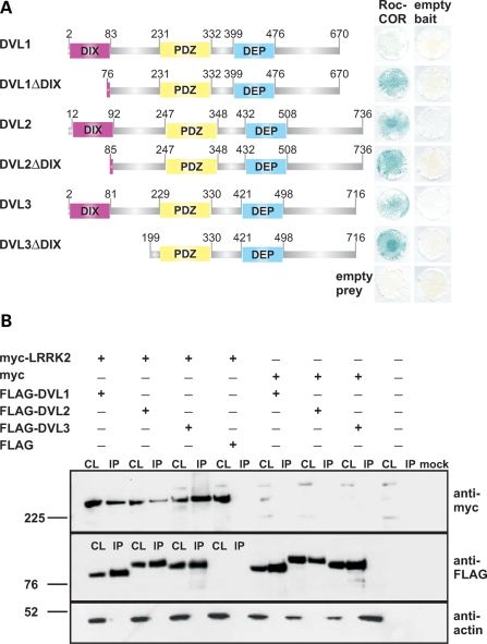
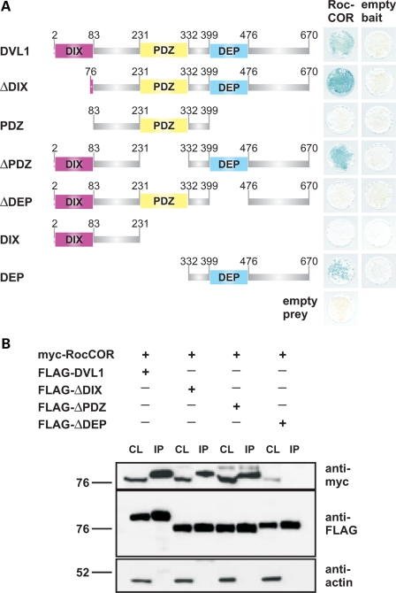
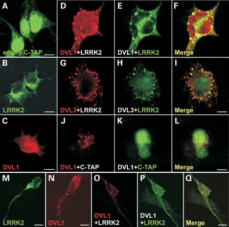
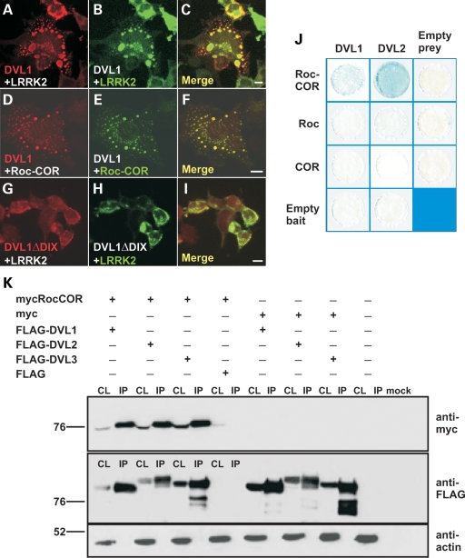
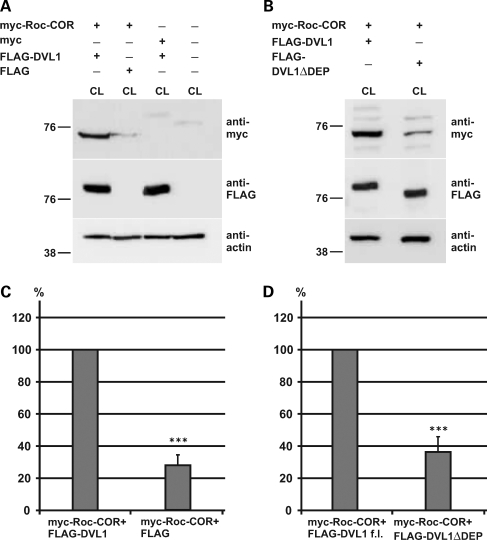
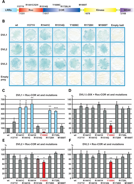
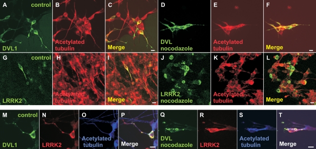
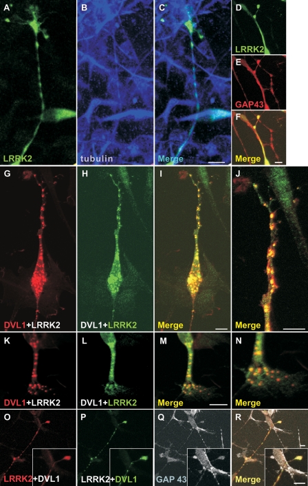
Mutations in PARK8, encoding LRRK2, are the most common known cause of Parkinson's disease. The LRRK2 Roc-COR tandem domain exhibits GTPase activity controlling LRRK2 kinase activity via an intramolecular process. We report the interaction of LRRK2 with the dishevelled family of phosphoproteins (DVL1-3), key regulators of Wnt (Wingless/Int) signalling pathways important for axon guidance, synapse formation and neuronal maintenance. Interestingly, DVLs can interact with and mediate the activation of small GTPases with structural similarity to the LRRK2 Roc domain. The LRRK2 Roc-COR domain and the DVL1 DEP domain were necessary and sufficient for LRRK2-DVL1 interaction. Co-expression of DVL1 increased LRRK2 steady-state protein levels, an effect that was dependent on the DEP domain. Strikingly, LRRK2-DVL1-3 interactions were disrupted by the familial PARK8 mutation Y1699C, whereas pathogenic mutations at residues R1441 and R1728 strengthened LRRK2-DVL1 interactions. Co-expression of DVL1 with LRRK2 in mammalian cells resulted in the redistribution of LRRK2 to typical cytoplasmic DVL1 aggregates in HEK293 and SH-SY5Y cells and co-localization in neurites and growth cones of differentiated dopaminergic SH-SY5Y cells. This is the first report of the modulation of a key LRRK2-accessory protein interaction by PARK8 Roc-COR domain mutations segregating with Parkinson's disease. Since the DVL1 DEP domain is known to be involved in the regulation of small GTPases, we propose that: (i) DVLs may influence LRRK2 GTPase activity, and (ii) Roc-COR domain mutations modulating LRRK2-DVL interactions indirectly influence kinase activity. Our findings also link LRRK2 to Wnt signalling pathways, suggesting novel pathogenic mechanisms and new targets for genetic analysis in Parkinson's disease.


Figures

Figure 1.
Figure 1.
The LRRK2 Roc-COR tandem domain interacts with dishevelled proteins DVL1, DVL2 and DVL3. (A) Full-length or partial DVL1, DVL2 and DVL3 preys were tested for interactions with the LRRK2 Roc-COR tandem domain bait in the YTH system using LacZ freeze-fracture assays. All negative controls show yeast growth but no blue colouration in the LacZ assay, demonstrating that the co-expression of bait and prey plasmids with empty prey or bait vectors does not result in transcription of reporter genes, i.e. no autoactivation was observed. Note that deletion of the DVL1 DIX domain (DVL1ΔDIX) increases the strength of the Roc-COR-DVL1 interaction. (B) Co-immunoprecipitation of LRRK2 and DVL1, DVL2 and DVL3 in HEK293 cells co-transfected with full-length myc-tagged LRRK2 and full-length FLAG-tagged DVL1, DVL2 or DVL3 constructs. Note that myc-LRRK2 is present in the cell lysates (CL) and FLAG-DVL1, FLAG-DVL2 and FLAG-DVL3 immunoprecipitation (IP) samples purified using FLAG beads, but not in IP samples from cells co-transfected with the empty FLAG vector.
Figure 2.
Figure 2.
The LRRK2 Roc-COR tandem domain interacts with the DVL1 DEP domain. (A) DVL1 deletion constructs lacking key domains (ΔDIX, ΔPDZ or ΔDEP), or encoding individual DIX, PDZ or DEP domains were tested for interactions with the LRRK2 Roc-COR tandem domain bait using the YTH system. LacZ freeze-fracture assays demonstrate that only constructs expressing the DVL1 DEP domain are able to interact with the Roc-COR bait, whereas the DIX and PDZ domains were dispensable. (B) Confirmation of the LRRK2-binding site on DVL1 by co-immunoprecipitation in HEK293 cells co-transfected with constructs encoding the myc-tagged LRRK2 Roc-COR tandem domain and FLAG-tagged DVL1 variants. Note that the myc-tagged LRRK2 Roc-COR tandem domain is present in the cell lysates (CL) and FLAG-DVL1, FLAG-DVL1ΔDIX, FLAG-DVL1ΔPDZ immunoprecipitation (IP) samples purified using FLAG beads, but not in IP samples from cells co-transfected with FLAG-DVL1ΔDEP.
Figure 3.
Figure 3.
Redistribution of LRRK2 to cytoplasmic aggregates containing DVL proteins and changes in cell morphology. Confocal microscopy showing the distributions of (A) TAP-epitope, (B) TAP-tagged full-length LRRK2 and (C) FLAG-tagged full-length DVL1. Note that the TAP epitope has a predominantly nuclear and cytoplasmic distribution, whereas TAP-LRRK2 is distributed evenly in the cytoplasm including cell processes. In contrast, FLAG-DVL1 is expressed in the cytoplasm forming round structures that represent DVL protein polymers (36). TAP-LRRK2 re-distributes to FLAG-DVL1 (DF) and FLAG-DVL3 (GI) aggregates upon co-transfection, and cells show unique changes in morphology, spreading and flattening out to cover a larger surface area. In contrast, the TAP epitope tag does not co-distribute with FLAG-DVL1 aggregates (JL), and changes in cell morphology are not observed. Similar results were obtained with myc-LRRK2, LRRK2-EGFP and LRRK2-V5-His (data not shown), suggesting that the redistribution of LRRK2 to DVL aggregates is independent on the nature of the epitope tag. A similar distribution of myc-LRRK2 (M) and FLAG-DVL1 (N) and the redistribution of myc-LRRK2 to FLAG-DVL1 aggregates (OQ) are seen in non-differentiated dopaminergic SH-SY5Y cells, demonstrating that this effect also occurs in different cell lines. Scale bars: 10 µm.
Figure 4.
Figure 4.
The cytoplasmic distribution of DVL–LRRK2 complexes is dependent on the DVL1 DIX domain and the intact LRRK2 Roc-COR tandem domain. Confocal microscopy showing the co-distribution of FLAG-tagged full-length DVL1 with myc-tagged full-length LRRK2 in cytoplasmic DVL protein polymers (AC). Since a myc-tagged Roc-COR construct shows similar targeting to cytoplasmic FLAG-DVL1 aggregates, the Roc-COR tandem domain is sufficient for this interaction (DF). Deleting the DIX domain by site-directed mutagenesis (DVL1ΔDIX) resulted in the loss of cytoplasmic DVL1 aggregates and the redistribution of FLAG-DVL1ΔDIX and myc-LRRK2 throughout the cytoplasm, but with apparent enrichment at the cell membrane (GI). Scale bars: 10 µm. (J) LacZ freeze-fracture assays demonstrate that the intact Roc-COR tandem domain, but not individual Roc or COR domains, binds DVL1 and DVL2 effectively. (K) Co-immunoprecipitation of the myc-tagged LRRK2 Roc-COR tandem domain by FLAG-tagged DVLs confirms that the LRRK2 Roc-COR tandem domain is sufficient for DVL binding. Note that the myc-tagged LRRK2 Roc-COR tandem domain is present in the cell lysates (CL) and enriched in FLAG-bead purified immunoprecipitation (IP) samples from co-transfections with FLAG-DVL1, FLAG-DVL2 and FLAG-DVL3, but not in control experiments using empty FLAG vector.
Figure 5.
Figure 5.
Co-expression of DVL1 stabilizes the expression of the LRRK2 Roc-COR tandem domain. (A) Co-expression of the myc-tagged Roc-COR tandem domain with FLAG-DVL1 results in a stabilization of the Roc-COR domain in cell lysates. (B) The stabilization of LRRK2 is dependent on the DVL1 DEP domain interaction with the Roc-COR tandem domain, since DVL1 lacking the interacting DEP domain (FLAG-DVL1ΔDEP) was not capable of stabilizing myc-Roc-COR. (C and D) Quantification of these effects after normalization to levels of cellular actin shows that FLAG-DVL1 increases myc-Roc-COR ∼4-fold compared with controls with an empty FLAG vector or FLAG-DVL1ΔDEP. Statistical significance was determined using a Student's t-test (two-tailed). Error bars represent the standard deviation of the mean. ***P < 0.001.
Figure 6.
Figure 6.
Modulation of the interaction between the LRRK2 Roc-COR tandem domain and dishevelled proteins (DVL1-3) by familial Parkinson's disease mutations. (A) Locations of amino acids affected by familial Parkinson's disease mutations in the Roc-COR tandem domain. (B) LacZ freeze-fracture assays of LRRK2-DVL interactions. Using this semi-quantitative assay, the most striking effect observed is that the Y1699C mutation in the COR domain clearly disrupts the LRRK2–DVL1 interaction. All negative controls show yeast growth but no blue colour in the LacZ assay, demonstrating that the co-expression of bait and prey plasmids with empty prey or empty bait vectors does not result in transcription of reporter genes, i.e. no autoactivation was observed. (CF) Quantitative liquid YTH assays using CPRG as substrate for β-galactosidase expression reveal that substitutions at R1441 and R1728 show a strengthened interaction between the Roc-COR domain bait and DVL1 prey, whereas Y1699C disrupted interactions between the Roc-COR domain bait and DVL1, DVL2 and DVL3 preys. Statistical significance was determined using a Student's t-test (two-tailed). Error bars represent the standard deviation of the mean. ***P < 0.001, **P < 0.01, *P < 0.05.
Figure 7.
Figure 7.
DVL1, but not LRRK2, stabilizes microtubules treated with nocodazole. (AL) Confocal microscopy showing stable microtubules stained for acetylated tubulin in SH-SY5Y cells 5 days after treatment with retinoic acid transfected with FLAG-DVL1 or myc-LRRK2. Note that in cells transfected with FLAG-DVL1, stable microtubules were observed both before (A–C) and after (D–F) nocodazole treatment. However, while acetylated tubulin was observed in cells expressing myc-LRRK2 before nocodazole treatment (G–I), LRRK2 alone was unable to protect microtubules from nocodazole treatment (J–L). Interestingly, cells co-transfected with FLAG-DVL1 and myc-LRRK2 showed stable microtubules both before (MP) and after (QT) nocodazole treatment. Scale bars: 10 µm.
Figure 8.
Figure 8.
Co-localization of LRRK2 and DVL1 in the cytoplasm, neurites and growth cones in differentiated dopaminergic cells. (AF) Confocal microscopy showing expression of myc-tagged LRRK2 in SH-SY5Y cells 5 days after treatment with retinoic acid. Note that LRRK2 co-localizes with tubulin staining in neurites (A–C) and GAP43 immunostaining in growth cones (D–F). Co-expression of FLAG-tagged DVL1 with myc-tagged LRRK2 in differentiated SH-SY5Y cells shows co-localization of both proteins in multiple cytoplasmic aggregates (GJ). (J) Magnification of the process shown in (G–I). Co-localization of LRRK2 and DVL1 in an extended neuronal terminal, suggestive of a growth cone (KN) and in growth cones co-stained for GAP43 (OR). Scale bars: 10 µm.

Similar articles

Cited by

References

    1. Paisán-Ruíz C., Jain S., Evans E.W., Gilks W.P., Simón J., van der Brug M., López de Munain A., Aparicio S., Gil A.M., Khan N., et al. Cloning of the gene containing mutations that cause PARK8-linked Parkinson's disease. Neuron. 2004;44:595–600. - PubMed
    1. Zimprich A., Biskup S., Leitner P., Lichtner P., Farrer M., Lincoln S., Kachergus J., Hulihan M., Uitti R.J., Calne D.B., et al. Mutations in LRRK2 cause autosomal-dominant parkinsonism with pleomorphic pathology. Neuron. 2004;44:601–607. - PubMed
    1. Healy D.G., Falchi M., O'Sullivan S.S., Bonifati V., Durr A., Bressman S., Brice A., Aasly J., Zabetian C.P., Goldwurm S., et al. Phenotype, genotype, and worldwide genetic penetrance of LRRK2-associated Parkinson's disease: a case–control study. Lancet Neurol. 2008;7:583–590. - PMC - PubMed
    1. Paisán-Ruíz C., Nath P., Washecka N., Gibbs J.R., Singleton A.B. Comprehensive analysis of LRRK2 in publicly available Parkinson's disease cases and neurologically normal controls. Hum. Mutat. 2008;29:485–490. - PubMed
    1. Khan N.L., Jain S., Lynch J.M., Pavese N., Abou-Sleiman P., Holton J.L., Healy D.G., Gilks W.P., Sweeney M.G., Ganguly M., et al. Mutations in the gene LRRK2 encoding dardarin (PARK8) cause familial Parkinson's disease: clinical, pathological, olfactory and functional imaging and genetic data. Brain. 2005;128:2786–2796. - PubMed

Publication types

MeSH terms

Substances