Comparative Study
. 2009 Nov;24(11):2767-77.
doi: 10.1093/humrep/dep247. Epub 2009 Jul 22.

L-selectin ligands in human endometrium: comparison of fertile and infertile subjects

Affiliations
Comparative Study

L-selectin ligands in human endometrium: comparison of fertile and infertile subjects

L Margarit et al. Hum Reprod. 2009 Nov.

Abstract

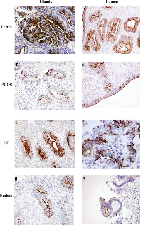
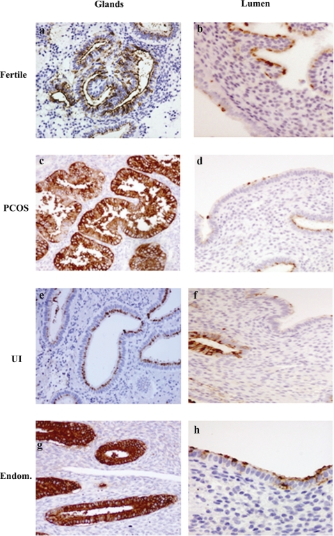
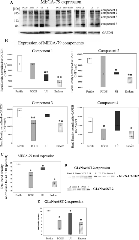
Background: L-selectin ligands, localized to the luminal epithelium at the time of implantation, may support the early stages of blastocyst attachment. We have assessed the expression of two L-selectin ligands, defined by MECA-79 and HECA-452 monoclonal antibodies, and the sulfotransferase GlcNAc6ST-2, involved in generation of L-selectin ligand epitopes, in the secretory phase of the endometrium from fertile and infertile patients.

Methods: Endometrial samples were obtained from 33 fertile, 26 PCOS, 25 endometriosis and 33 patients diagnosed with unexplained infertility. L-selectin ligands and GlcNAc6ST-2 expression was assessed by immunohistochemistry and immunoblotting.

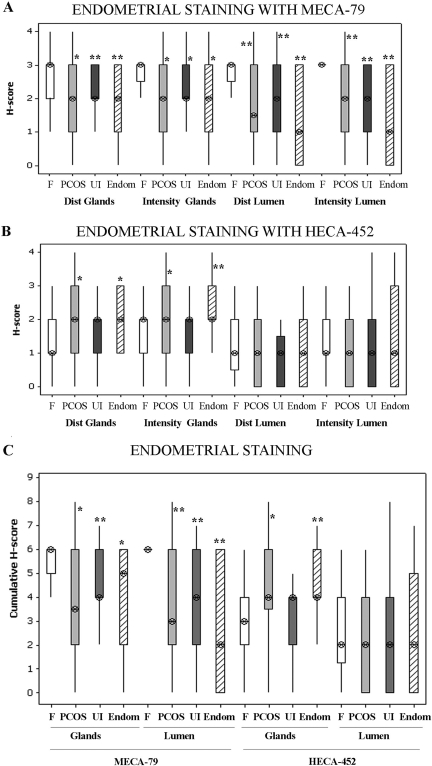
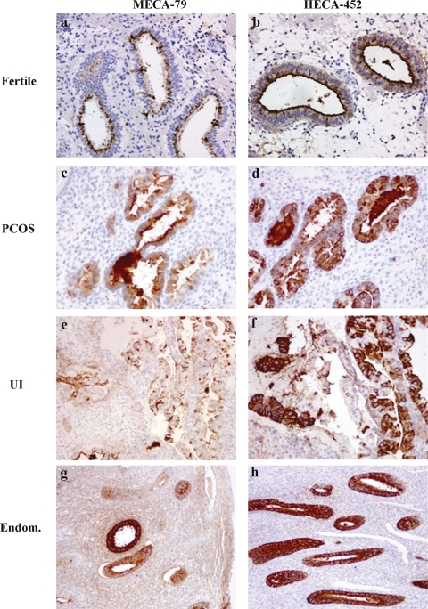
Results: Immunohistochemical staining of uterine epithelium, from fertile and infertile women, demonstrated differential expression of MECA-79 and HECA-452 epitopes. In fertile women in the secretory phase MECA-79 was more strongly expressed, particularly on the lumen, than in infertile women. HECA-452 staining was significantly stronger in the glands in PCOS and endometriosis patients than in fertile women. GlcNAc6ST-2 expression was reduced in infertile patients, correlating with MECA-79 expression.

Conclusions: This study demonstrated significant differences in expression of L-selectin ligands between fertile and infertile women in natural cycles, and could contribute to patient assessment prior to initiating fertility treatment.


Figures

Figure 1
Figure 1
Expression of MECA-79 in glands and lumen in the secretory phase endometrium. Secretory phase endometrium from fertile (33 patients), endometriosis (endom) (25 patients), PCOS (26 patients) and unexplained infertility (UI) patients (33 patients) was analyzed for the expression of MECA-79 as described in Materials and Methods. In fertile women MECA-79 was strongly expressed on the luminal epithelium and the glands compared with endometriosis (endom.), PCOS and UI . Magnifications ×400 (a, d, e and f), ×200 (b, c, g and h). Statistical analysis of the immunohistochemistry scores is showed in Fig. 3A.
Figure 2
Figure 2
Expression of HECA-452 in glands and lumen in the secretory phase endometrium. Secretory phase endometrium from fertile (33 patients), endometriosis (endom) (25 patients), PCOS (26 patients) and unexplained infertility (UI) patients (33 patients) was analyzed for the expression of HECA 452 as described in Materials and Methods. In endometriosis (endom.) and PCOS the staining in the glands was significantly stronger that in the fertile group. The UI group did not present any significant difference in staining of the glands compared with the fertile group (see Fig. 3B). Magnifications ×400 (b, h), ×200 (a, c, d, e, f and g). Statistical analysis of the immunohistochemistry scores is showed in Fig. 3B.
Figure 3
Figure 3
MECA-79 and HECA-452 expression, intensity and distribution of staining in lumen and glands. (A) and (B) Intensity and distribution scores for staining of HECA and MECA antibodies in endometrium of fertile and study groups are plotted. (C) Cumulative scores for each antibody and groups are plotted. Values are median (⊗) and inter-quartile range (box and whisker). The Kruskal–Wallis test followed by a Mann–Whitney test was performed to determine first statistical significance between groups and secondly to compare specific group pairs. *P ≤ 0.05 and **P ≤ 0.01 are considered significant and are compared with fertile patients. Fertile (white box, n= 33 patients), PCOS (grey box, n = 26 patients), UI (Dark grey box, n = 33 patients), Endometriosis (Endom, hatched box, n = 25 patients).
Figure 4
Figure 4
Relation between the staining with HECA-452 and MECA-79 in endometrium of fertile and infertile women. Same regions were compared for staining with HECA-452 and MECA-79 for all groups. In all cases a reciprocal relation between staining with MECA-79 and HECA-452 was observed regardless of the fertility status of the patients. Fertile (n = 33 patients), PCOS (n = 26 patients), UI (n = 33 patients), Endometriosis (Endom, n = 25 patients). Magnifications ×200 (a, b, c, d and f) ×100 (e, g and h).
Figure 5
Figure 5
Expression of MECA-79 and sulfotransferase (GlcNAc-GST2) in epithelial cells isolated from endometrial biopsies. (A) and (D) Protein extracts of epithelial cells isolated from biopsies obtained from fertile (F, n = 6), endometriosis (Endom or Endo, n = 6), PCOS (n = 6) and UI (n = 6) patients were analyzed for the expression of MECA-79 (A) and GlcNAc-GST2 (D) as described in Materials and Methods. Protein levels were normalized to GAPDH protein levels (A). In fertile (F) women MECA-79 as well as GlcNAc-GST2 was strongly expressed compared with endometriosis (endom.), PCOS and UI. Representative blots are showed. (B) Densitometric analysis of each MECA-79 component was performed using the Quantity One software (Bio-Rad). Values are band density normalized to GAPDH and are expressed as intensity per square milimiters (INT*mm2). Values are expressed as median (⊗) and inter-quartile range (box and whisker). Fertile (white box), PCOS (grey box), UI (Dark grey box), Endometriosis (Endom, hatched box). (C) Densitometric analysis of the area covering all MECA-79 components was perfomed using the Quantity One software (Bio-Rad). Values are band density normalized to GAPDH and expressed as Intensity per square milimiters (INT*mm2). Values as expressed as median (⊗) and inter-quartile range (box and whisker). Fertile (white box), PCOS (grey box), UI (Dark grey box), Endometriosis (Endom, hatched box). (E) Densitometry analysis of the immunoblots GlcNAc-GST2 levels was performed using the Quantity One software (Bio-Rad). Values obtained are expressed as band density compared with fertile patients and are expressed as percent. Values are median (⊗) and inter-quartile range (box and whisker). Fertile (white box), PCOS (grey box), UI (Dark grey box), Endometriosis (Endom, hatched box). Statistical analysis of the samples (B, C and E) was performed using the Kruskal–Wallis test to determine significance between groups followed by a Mann–Whitney test to compare specific group pairs. *P ≤ 0.05 and **P ≤ 0.01 are considered significant.

Similar articles

Cited by

References

    1. Alon R, Feigelson S. From rolling to arrest on blood vessels: leukocyte tap dancing on endothelial integrin ligands and chemokines at sub-second contacts. Semin Immunol. 2002;14:93–104. - PubMed
    1. Ben-Nun I, Jaffe R, Fejgin MD, Beyth Y. Therapeutic maturation of endometrium in in vitro fertilization and embryo transfer. Fertil Steril. 1992;57:953–962. - PubMed
    1. Bistrup A, Tsay D, Shenoy P, Singer MS, Bangia N, Luther SA, Cyster JG, Ruddle NH, Rosen SD. Detection of a sulfotransferase (HEC-GlcNAc6ST) in high endothelial venules of lymph nodes and in high endothelial venule-like vessels within ectopic lymphoid aggregates: relationship to the MECA-79 Epitope. Am J Pathol. 2004;164:1635–1644. - PMC - PubMed
    1. Bos J, de Boer O, Tibosch E, Das P, Pals S. Skin-homing T lymphocytes: detection of cutaneous lymphocyte-associated antigen (CLA) by HECA-452 in normal human skin. Arch Dermatol Res. 1993;285:179–183. - PubMed
    1. Carson DD, Bagchi I, Dey SK, Enders AC, Fazleabas AT, Lessey BA, Yoshinaga K. Embryo implantation. Dev Biol. 2000;223:217–237. - PubMed

Publication types